Shed Exoskeleton of a Spider and its Leg Hair
 Nov 05, 2020 • 10:41 PM UTC
 Unknown Location
 140x Magnification
 Unknown
Elton Tran
Learn about the author...
1posts
0comments
1locations
View in Media Gallery
In terms of diversity of insects and organisms that can be visualized under a Foldscope slide, the Sacramento suburbs don’t seem immediately to offer too much beyond ants and moss. I didn’t have high hopes when I went outside to my backyard in an attempt to find something exciting to mount and observe. I settled on collecting spider web samples to mount, but I didn’t notice until later that I unknowingly also collected a souvenir from the web’s past occupant: a relatively intact shed spider exoskeleton.
View in Media Gallery
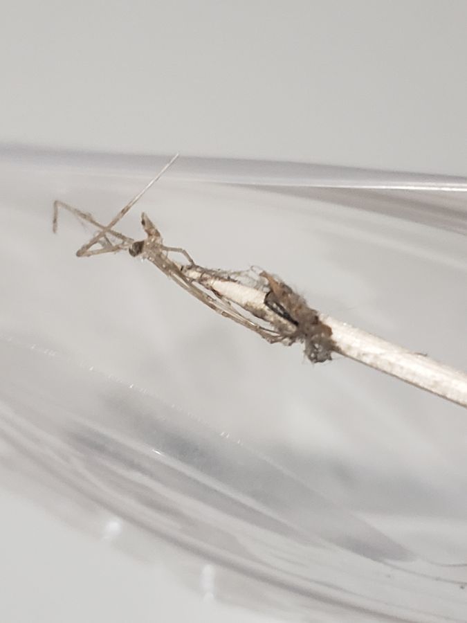
Shed exoskeleton along with the original web sample I was immediately happy to switch gears. It’s difficult to tell from the exoskeleton what type of species of spider it belonged to, but looking at the sample underneath the Foldscope allowed me to see the parts of the spider’s outer anatomy that were preserved by the exoskeleton.
View in Media Gallery
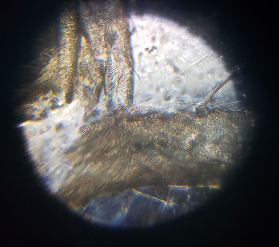
At this angle, I was able to see the break in the molt at a leg segment where the spider seemed to have emerged from. What was interesting to see was that there were hairs that conspicuously grew from the spider’s leg, which weren’t noticeable or observable at all without magnification. Spiders rely on hair follicles as sensory anatomy, so the presence of hair in the shed exoskeleton was a surprising finding for me. Are hairs grown underneath an exoskeleton that’s ready to shed and are ready to use right after molting? Or do spiders have to go through a period of vulnerability with reduced sensory ability by regrowing their hairs after shedding? Looking at the anatomy of the spider molt certainly brought up new questions about spider physiology and how molting affects their ability to continue about their lives.
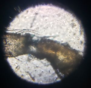
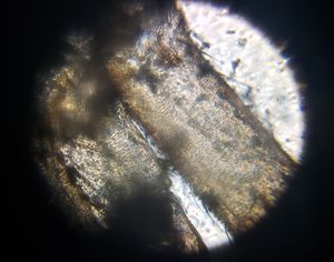
Above, you can see different parts of the exoskeleton that I was able to clearly observe. The hairs are again conspicuous on the left photo, but on a leg segment that wasn’t broken in the process of molting. On the right is where two leg segments connected to the spider’s main body, with its transparency revealing that its past owner had definitely left this molt behind for good.
I was glad to get a chance to personally take a closer look at the previously invisible parts of a spider’s anatomy.
I conducted this project as part of Professor Pringle’s EEB321 class at Princeton University.
I was glad to get a chance to personally take a closer look at the previously invisible parts of a spider’s anatomy.
I conducted this project as part of Professor Pringle’s EEB321 class at Princeton University.
Sign in to commentNobody has commented yet... Share your thoughts with the author and start the discussion!
More Posts from Elton Tran
No more posts from this author.