Homemade Mosquito Larvae
 Nov 09, 2020 • 10:07 AM UTC
 Unknown Location
 140x Magnification
 Unknown
Janet You
Learn about the author...
1posts
0comments
1locations
View in Media Gallery
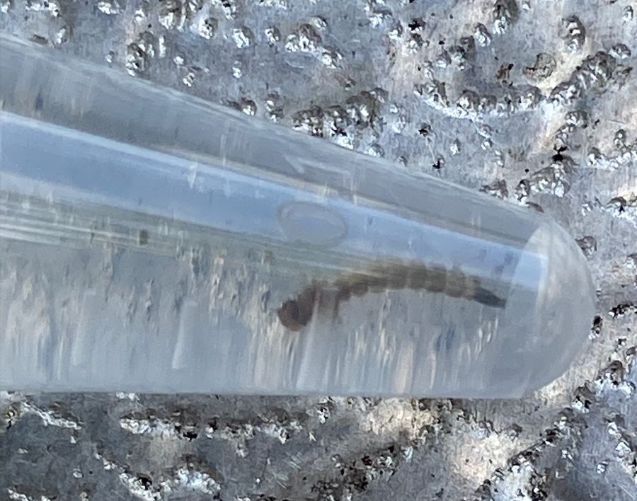
After receiving my foldscope in the mail, I was incredibly excited on having my own at-home, usable microscope. I was so excited to be grabbing samples from everywhere in my house. I tried to think of something I really wanted to look at under a microscope and then it hit me. This past summer, I was working with mosquitos for my senior thesis project. During this data collection period, I actually spent quite a lot of time collecting mosquito larvae and pupae (to grow into adults in tubes) so I can identify them once I return to the lab on campus. Unfortunately, many of the larvae and pupae I collected don’t survive into the adult phase in the collection tubes.
View in Media Gallery
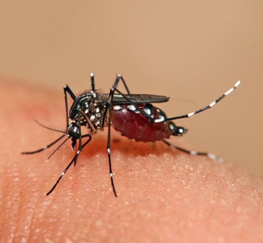
Aedes mosquito I wanted to take a microscopic look at one of these dead larvae. After all, “ecology is everywhere” and this mosquito larvae was taken straight from a container in my backyard. I made sample with the larvae and took a look.
View in Media Gallery
A larvae I collected over the summer I was interested if the microscopic image help me to identify the species of the mosquito. I had hypothesized that the mosquito was an Aedes mosquito rather than a Culex mosquito due to the tail shape. However, under the lens, I felt it was actually harder to tell due to the close up nature of the image.
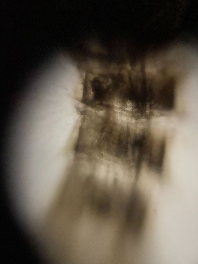
View in Media Gallery
Foldscope view It was really shocking to see how transparent the larvae. In person, the larvae gives off this really dark (almost black) color. It is also really interesting to see the different sections. What is the benefit of this structure of their body for mosquito larvae? Why is their body sectioned? How does larvae develop into pupae and what physical changes does the larvae go through when it becomes a pupa. Some larvae were bigger than other ones, so how is this size difference reflected under the microscope?
I was in the fortunate situation of having this opportunity to be able to view mosquito larvae under the foldscope. It was amazing to see how well the foldscope works and to have a microscopic view of something I have become so familiar with during the summer. It was super fascinating to see how the microscopic view looked so different than what I see in the tube and how it revealed certain qualities of the larvae I would not have caught with the human eye. I would definitely use the foldscope again and test it on other items to see what it could reveal that the human eye can’t capture.
I conducted this project as part of Professor Pringle’s EEB321 class at Princeton University.
Sign in to commentNobody has commented yet... Share your thoughts with the author and start the discussion!
More Posts from Janet You
No more posts from this author.