Stomata and Guard Cells
 Nov 27, 2020 • 2:47 AM UTC
 Unknown Location
 140x Magnification
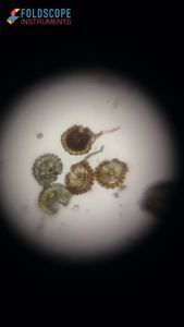
 Microorganisms
Diya A
Learn about the author...
13posts
9comments
1locations
View in Media Gallery
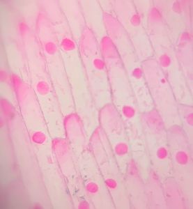
I observed the outer epidermis of a leaf and the skin of a pea pod under my Foldscope. The samples were taken at different times of the day, and we can see both closed and open stomata, depending on when they were taken.
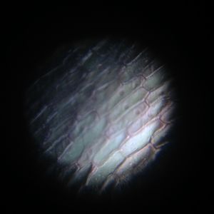
Closed Stomata
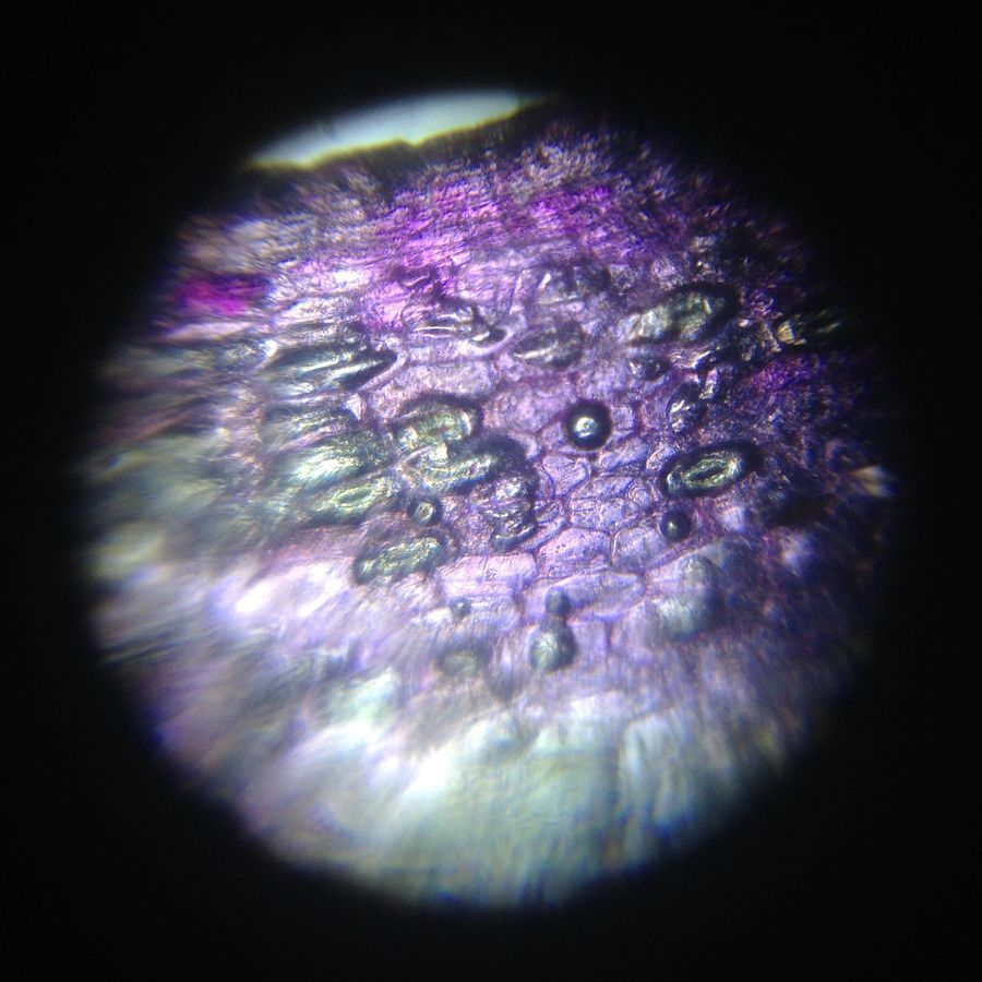
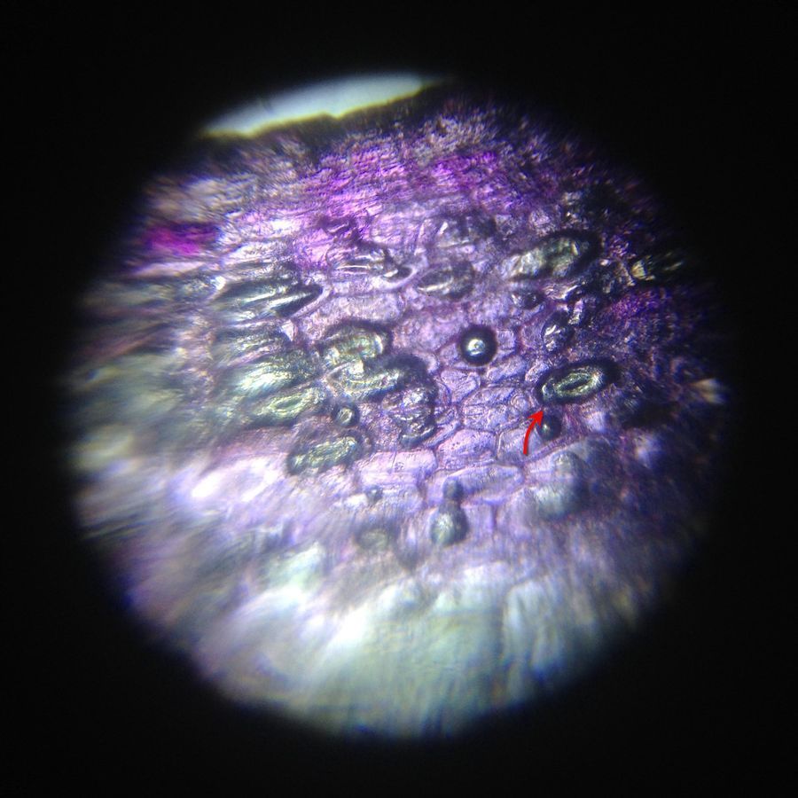
This is a leaf peel from a rhoeo ( Tradescantia spathacea ) plant. This plant’s leaves are generally observed in an introduction to school-level microscopy. The sample has been taken late in the evening.
Closed Stomata
This is a leaf peel from a rhoeo ( Tradescantia spathacea ) plant. This plant’s leaves are generally observed in an introduction to school-level microscopy. The sample has been taken late in the evening.
View in Media Gallery
The stomata are pores scattered among the cells, and are closed. A pair of guard cells surrounds each pore.
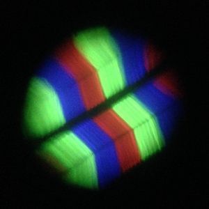
Open Stomata
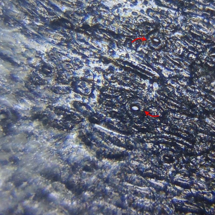
This is the skin of a fresh green pea pod. The sample has been taken during the daytime, around midday.
Open Stomata
This is the skin of a fresh green pea pod. The sample has been taken during the daytime, around midday.
View in Media Gallery
Here, the stomata are wide open. Kidney bean-shaped guard cells are seen around the pores.
CookerBird
CookerBird
Sign in to commentNobody has commented yet... Share your thoughts with the author and start the discussion!

 0 Applause
 0 Comments