I. Understanding the earliest farmers: The “crops”
 Apr 02, 2021 • 11:46 PM UTC
 Unknown Location
 140x Magnification
 Microorganisms
Laks Iyer
Human observer of life. https://sukshmadarshin.wordpress.com
97posts
1255comments
5locations
View in Media Gallery
Lockdown and working from home have their own advantages (let us only talk about good things). My favorite new pastime is interacting with an 11-year-old and her everyday school lessons, sometimes even as the class is done. A few days ago, the 11-year-old lectured me on the earliest farmers in the fertile crescent, the Indian subcontinent, and the Far East. She laid the current model of the history of crop and animal domestication on a large map on our table and the transition from hunter-gatherers to the earliest aggregates of large populations. I relearned that farming and domestication from 10-15,000 years ago was a major transition in human society. Like in my school classrooms, I began day-dreaming the idea of domestication and suddenly emerged in the discussion a trick question—- are we, humans, the first farmers/domesticators?
I am sure you know that NO is the answer. It turns out that we are late in this game of farming/domestication for food. Studies show that farming is widespread among invertebrates–Several ant species, termites, beetles, and even the marine snail Littoraria, all of whom preceded us by over 50 million years, farm fungi. Ants tend to aphids for their honeydew. A more interesting type of farming is seen in the social amoeba Dictyostelium. They carry their food as they are dispersed as spores, giving them a ready food source in nutrition/bacteria-poor regions. Some of the fascinating associations are those of the Lichens, made of fungi collaborating with photosynthetic bacteria or algae. I am really not sure who is farming whom in this case. Here I wish to lay down some thoughts on the farming of photosynthetic life.
The evolution of photosynthesis; Carbon-fixation using light as an energy source was a big game-changer in the history of life on earth. About 2 billion years ago, there was massive oxygenation of the earth’s atmosphere. Evidence suggests that the cyanobacteria drove this process probably due to the evolution of photosynthesis in them. Part I of this series of posts is about some of the cyanobacteria I have studied recently. Colloquially referred to as Blue-green algae, these are found almost anywhere around us. Unlike most bacterial groups, they came in attractive multicellular aggregates and may often have specialized cells. The fascinating thing is that cyanobacteria are some of the earliest “crops” that have been domesticated over and over again and that is the essence of this series.
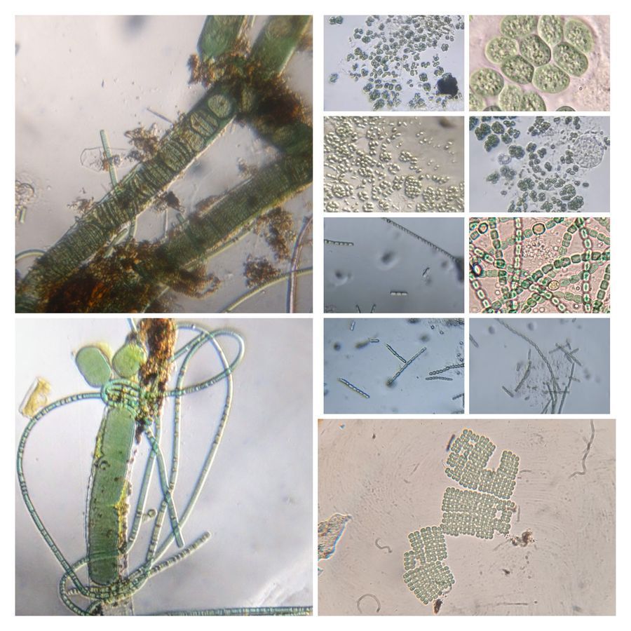
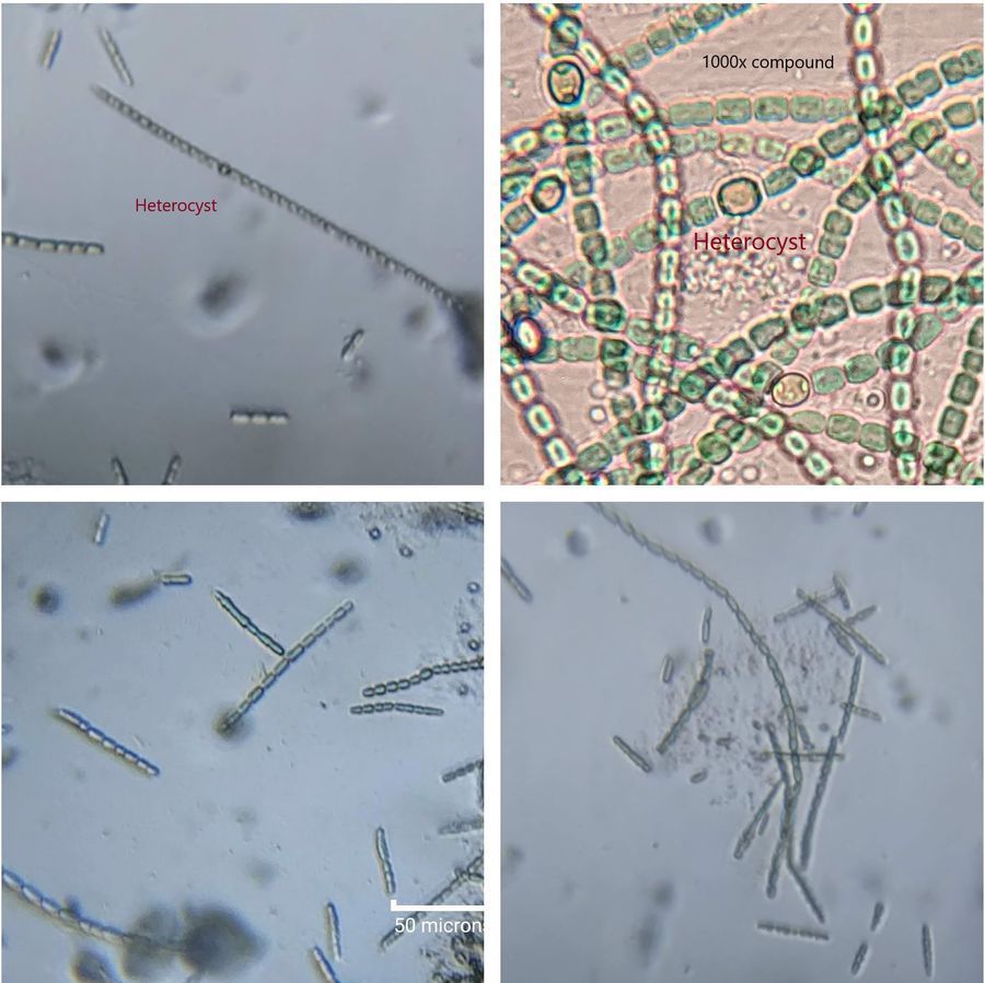
In the next picture is the Cyanobacterium Anabaena or Nostoc . Notice that these cells are in chains (about 3-4 microns is the cell length). If you look carefully, these chains are interrupted by globular cells called heterocysts. I have included a higher magnification compound microscope image of the same. This specialized cell creates an anaerobic environment for fixing Nitrogen, i.e. converting atmospheric nitrogen into an organic form such as ammonia and its derivatives. This process is key for converting atmospheric nitrogen to a form usable for the rest of us. Here is a fun fact. This culture of mine I first obtained in May 2020. I left it in the same tube and today, April 2021, it still seems to be growing well. Photosynthesis provides a tremendous survival advantage. Nostoc , Anabaena and other cyanobacteria can also completely pollute water bodies to give algal blooms and often release serious toxins in the waters. Recently, a large number of elephants were killed in Botswana near water bodies, and cyanobacterial toxins are proposed to be the main culprit ( Read here ).
I am sure you know that NO is the answer. It turns out that we are late in this game of farming/domestication for food. Studies show that farming is widespread among invertebrates–Several ant species, termites, beetles, and even the marine snail Littoraria, all of whom preceded us by over 50 million years, farm fungi. Ants tend to aphids for their honeydew. A more interesting type of farming is seen in the social amoeba Dictyostelium. They carry their food as they are dispersed as spores, giving them a ready food source in nutrition/bacteria-poor regions. Some of the fascinating associations are those of the Lichens, made of fungi collaborating with photosynthetic bacteria or algae. I am really not sure who is farming whom in this case. Here I wish to lay down some thoughts on the farming of photosynthetic life.
The evolution of photosynthesis; Carbon-fixation using light as an energy source was a big game-changer in the history of life on earth. About 2 billion years ago, there was massive oxygenation of the earth’s atmosphere. Evidence suggests that the cyanobacteria drove this process probably due to the evolution of photosynthesis in them. Part I of this series of posts is about some of the cyanobacteria I have studied recently. Colloquially referred to as Blue-green algae, these are found almost anywhere around us. Unlike most bacterial groups, they came in attractive multicellular aggregates and may often have specialized cells. The fascinating thing is that cyanobacteria are some of the earliest “crops” that have been domesticated over and over again and that is the essence of this series.
In the next picture is the Cyanobacterium Anabaena or Nostoc . Notice that these cells are in chains (about 3-4 microns is the cell length). If you look carefully, these chains are interrupted by globular cells called heterocysts. I have included a higher magnification compound microscope image of the same. This specialized cell creates an anaerobic environment for fixing Nitrogen, i.e. converting atmospheric nitrogen into an organic form such as ammonia and its derivatives. This process is key for converting atmospheric nitrogen to a form usable for the rest of us. Here is a fun fact. This culture of mine I first obtained in May 2020. I left it in the same tube and today, April 2021, it still seems to be growing well. Photosynthesis provides a tremendous survival advantage. Nostoc , Anabaena and other cyanobacteria can also completely pollute water bodies to give algal blooms and often release serious toxins in the waters. Recently, a large number of elephants were killed in Botswana near water bodies, and cyanobacterial toxins are proposed to be the main culprit ( Read here ).
View in Media Gallery
The cyanobacterium Merismopedia is the hero of the following movie. These cyanobacteria are arranged in a grid and held in a mucilagenous matrix. Inspired by previous posts in microcosmos, I made a timelapse of Merismopedia motion. It is fascinating to see how they move. Unlike many bacteria, cyanobacteria lack flagella, and their motility is supposed to involve a structure known as the pilus, and mucilage is constantly secreted as a medium for the movement, or colloquially a medium to slip on.
Next is the cyanobacterium Gleocapsa . Here you can see recently divided cells enclosed in a gelatinous capsule. They are usually clumped together as seen. I have also included a compound microscope view of them at 1000x under oil immersion. Early studies in Gleocapsa had shown that they also fix nitrogen independent of a heterocyst-like structure. Perhaps the capsule around them gives them an anaerobic environment.

View in Media Gallery
Gleocapsa under Foldscope. The compound microscope image is also shown Whereas some cyanobacteria are extremely toxic, others such as Spirulina are used as food sources. I prefer watching Spirulina under the microscope over eating them as chips. I was expecting a very visual spiral organism from this pure culture, but perhaps one needs to look at them under a compound microscope to see the Spiral shape. These do move really fast. Here is a timelapse of a Spirulina sample taken for 30 minutes with a picture every 10 seconds
Next is an Oscillatoria or its relative gliding through my foldscope. The water sample is from a wonderful place called Adyar Tolkappiar Poonga very close to my ancestral home. It is remarkable in that the wildlife recreates what I had seen in my childhood around our ancestral home,
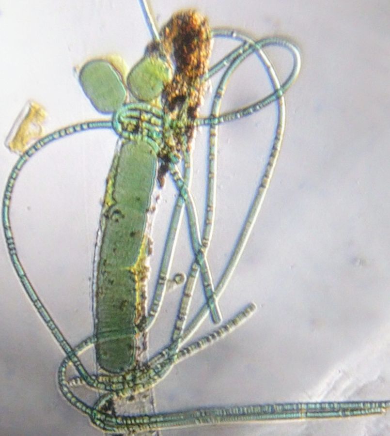
Also found in the Poonga was this wonderful clump of cyanobacteria that combines a broad Lynbia filament with a thin Phormidium filament. I thought it looked like an L for Leeuwenhoek, one of my many heroes. The crowning glory of this picture was that it was accepted as a cover image for one of my research papers (I am otherwise a theoretician) and it made me immensely proud ( Read this ). For those interested in the evolution of bacterial multicellularity and the evolution of apoptosis, there is a lot of interesting discovery in the paper. In Part II of my post, I shall describe how cyanobacteria are at the base of some of the oldest domestications in the history of life.
View in Media Gallery
Sign in to commentNobody has commented yet... Share your thoughts with the author and start the discussion!

 0 Applause
 0 Comments
_300x300.jpeg)