Sedum adolphi ‘Firestorm’
 Dec 15, 2020 • 11:25 AM UTC
 Unknown Location
 140x Magnification
 Microorganisms
Frances Phung
Learn about the author...
1posts
0comments
1locations
View in Media Gallery
As a succulent enthusiast, the object I have chosen for scientific curiosity is a succulent called Sedum adolphi ‘firestorm,’ which were taken from my personal collection of succulents. I find it exciting to learn about the utilization organisms to produce or carry out functions related to the biomedical field. Examples are Escherichia coli , Saccharomyces cerevisiae , or yeast to produce insulin or viral vectors to deliver gene therapy. After some time in my undergraduate program, I developed an appreciation for the solutions to human health that nature can offer. As a biomedical engineer, I only had the desire to focus on animal cells and the mammalian system; I want to broaden my scope and open my eyes to things around me that can be studied and used improve medicine. That is why I took a sample from one of my beloved succulents. To prepare these images, I tried to skin the stem and the leaf and thin as I could manage as I assumed the Foldscope would give results similar to a compound light microscope. My first question was whether I could see chloroplasts with the Foldscope, and my second was merely whether I can create mathematical models of plant cells to make them analogous to circuits just as how animal cells and the human body can. Luckily, with samples of the right thickness I was able to clearly see chloroplasts! As I partially skinned the bottom of a succulent leaf, I think I have identified two stomata! As the second question I asked is an open ended one, I have no conclusion other than to remember to broaden my scope for learning and to appreciate the potential nature has in future biomedical engineering research.
View in Media Gallery
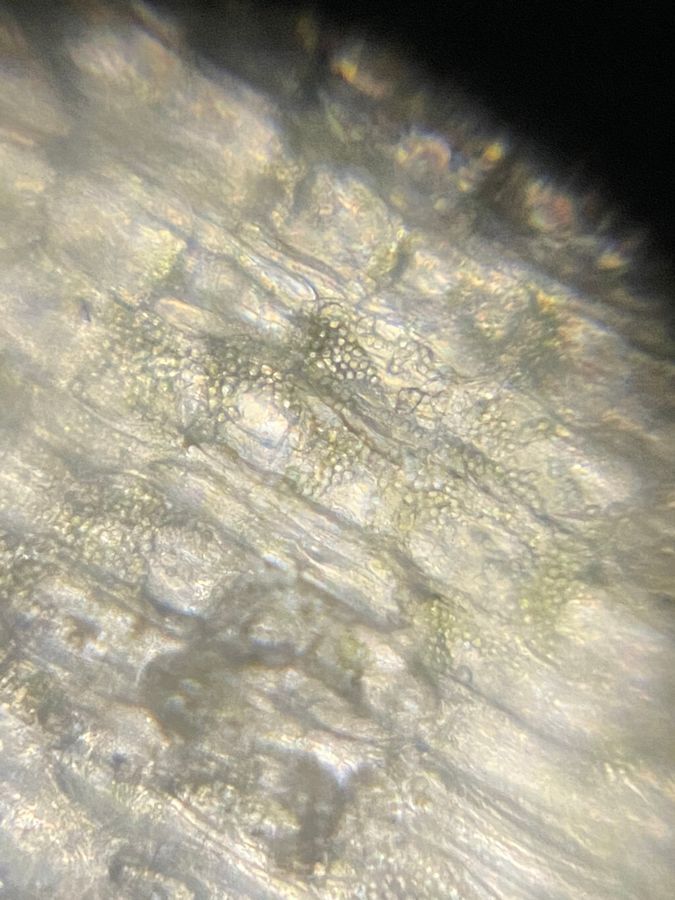
Figure 1. A thin, skin sample of the stem of S. adolphi ‘firestorm’ seen with a foldscope and a light source and taken with an iPhone11 Pro Max at the 1x setting. Characteristics easily observed are the plant cell walls, chloroplasts, and full vacuoles.
View in Media Gallery
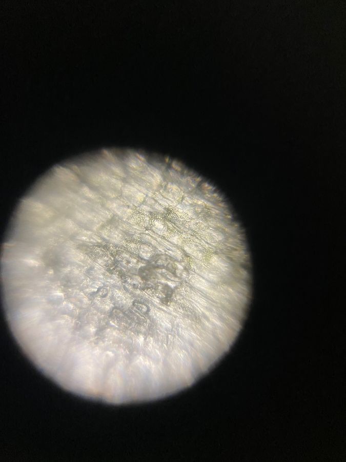
Figure 2.. A thin, skin sample of the stem of S. adolphi ‘firestorm.’ Characteristics easily observed are the plant cell walls, chloroplasts, and full vacuoles. The foldscope image remained the same; an iPhone 11 Pro Max was used to zoom in on the defined features for a better visualization.
View in Media Gallery
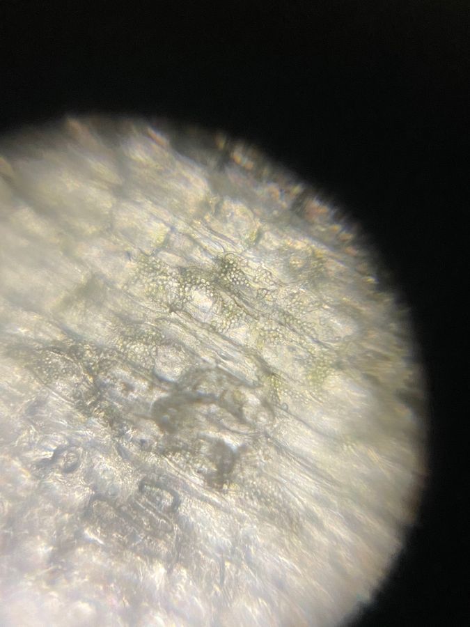
Figure 3. A thin, skin sample of the stem of S. adolphi ‘firestorm.’ Characteristics easily observed are the plant cell walls, chloroplasts, and full vacuoles. The foldscope image remained the same; an iPhone 11 Pro Max was used to further zoom in on the defined features for a better visualization.
View in Media Gallery
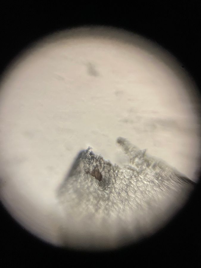
Figure 4. A thin, skin sample of a leaf of S. adolphi ‘firestorm.’ Characteristics easily observed are the plant cell walls and full vacuoles. An iPhone 11 Pro Max was used to take the photo and zoom in on the defined features. I believe there are two stomata toward the center of the image.
Sign in to commentNobody has commented yet... Share your thoughts with the author and start the discussion!
More Posts from Frances Phung
No more posts from this author.