Spores, pollen, wonder and more
 Apr 22, 2021 • 12:23 PM UTC
 Unknown Location
 140x Magnification
 Microorganisms
Diya A
Learn about the author...
13posts
9comments
1locations
View in Media Gallery
This is a documentation of two intended samples and one hitch-hiked sample, all very different from each other yet peacefully inhabiting the same square foot habitat. Plus one bonus sample at the end!
When I first learnt about fern spores, I was intrigued by how an entire plant grew out of a microscopic grain, and not a macroscopic seed. I went to ask a neighbour for a fern leaf to try and discern the spores, but the variety she had was “spore-less”. A year or two later I came across the word “sporange” (the only word that rhymes with “orange”), a shortened version of “sporangium”.
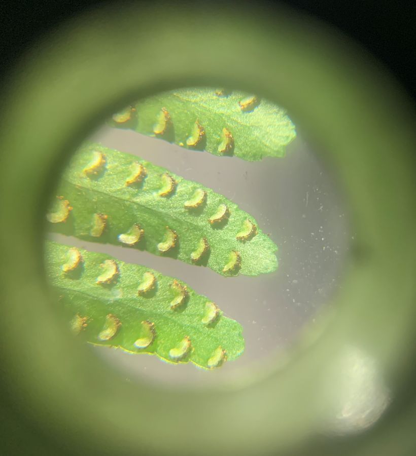
Yesterday I chanced upon a few ferns, and it kindled my curiosity. When I observed the leaf, I saw green semicircular structures on its underside, and brown powdery grains on the curved part, which I assumed to be the sporangia and spores respectively.
When I first learnt about fern spores, I was intrigued by how an entire plant grew out of a microscopic grain, and not a macroscopic seed. I went to ask a neighbour for a fern leaf to try and discern the spores, but the variety she had was “spore-less”. A year or two later I came across the word “sporange” (the only word that rhymes with “orange”), a shortened version of “sporangium”.
Yesterday I chanced upon a few ferns, and it kindled my curiosity. When I observed the leaf, I saw green semicircular structures on its underside, and brown powdery grains on the curved part, which I assumed to be the sporangia and spores respectively.
View in Media Gallery
Underside of fern leaf under 20x magnification
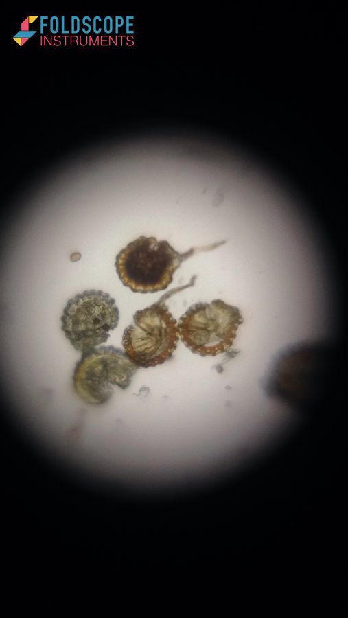
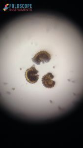
But under the Foldscope, I observed the “spores” bursting open, and releasing tinier “sporettes”! So I did a bit of research, and found out that the semicircular structures weren’t sporangia, but sori (singular sorus), and the brown powder grains were sporangia. The “sporettes” were actually the spores.
Here is a time-lapse, though the process was quite fast and I should have taken a regular video instead.
View in Media Gallery
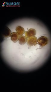
Sporangia releasing spores
View in Media Gallery
Some sporangia were empty and didn’t burst
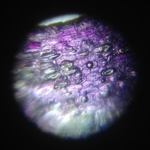
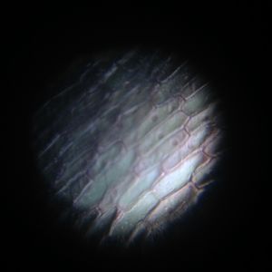
Then I observed an entire sorus. Here are different parts in focus; I tried image stitching, but it really didn’t work out for this one.
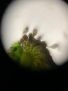
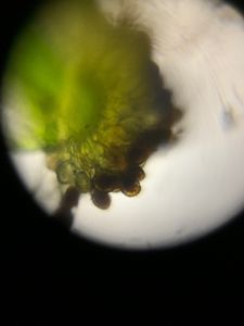
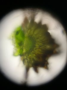
The green part of the sorus looks like it is made of immature green sporangia
The second sample is a beautiful white bougainvillea flower that had fallen under the fern.
The second sample is a beautiful white bougainvillea flower that had fallen under the fern.
View in Media Gallery
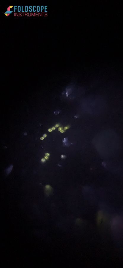
I observed a bract first, but it wasn’t anything unique. Next was the bright yellow pollen, hiding inside the stem of the little white flowers.
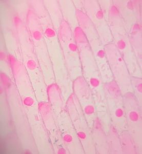
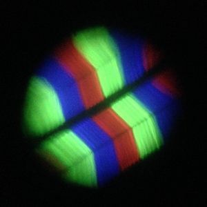
I’ve never seen anything like these pollen grains! They were extremely tiny. And they looked just like yellow RBCs, with a characteristic biconcave shape!
I’ve never seen anything like these pollen grains! They were extremely tiny. And they looked just like yellow RBCs, with a characteristic biconcave shape!
View in Media Gallery
Perhaps the biconcave shape maximizes surface area – but why?
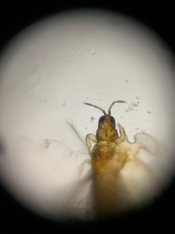
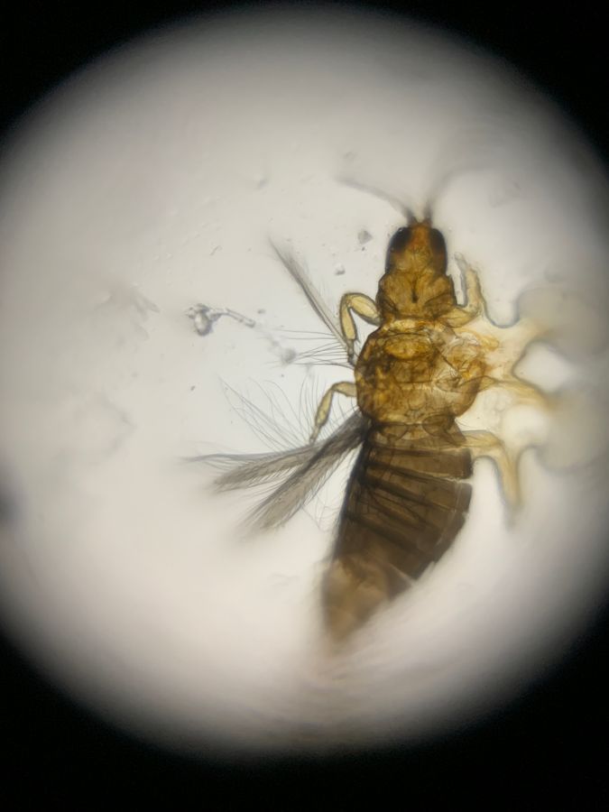
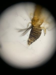
The next sample was a very small insect that rode along with the leaf and flower. It was glinting black to the unaided eye, but brown and translucent under magnification!
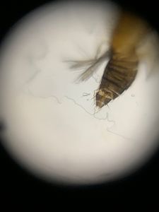
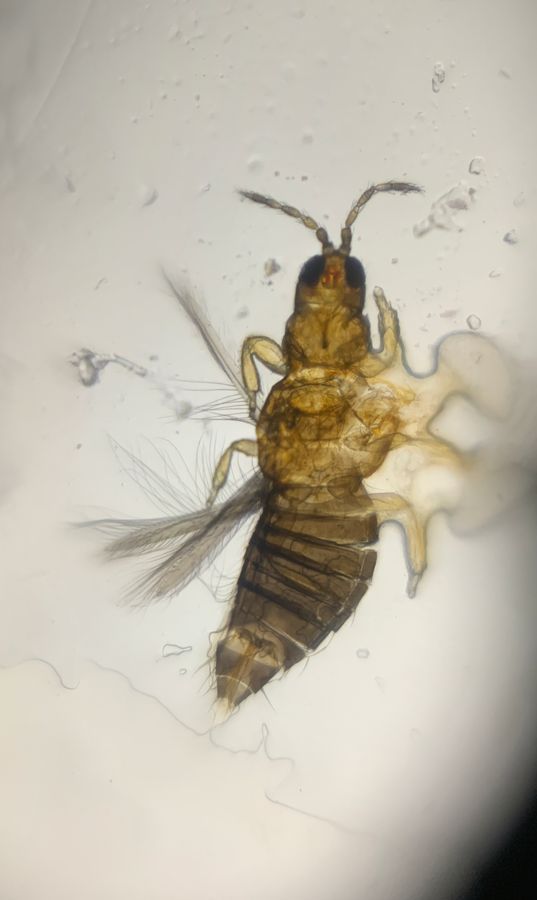
Here are photos of the bug from top to bottom in focus.
The next sample was a very small insect that rode along with the leaf and flower. It was glinting black to the unaided eye, but brown and translucent under magnification!
Here are photos of the bug from top to bottom in focus.
View in Media Gallery
Could that little red thing between its eyes be its brain?
View in Media Gallery
I tried not to, but (eek!) the poor little thing got squished
Does anyone know what insect this could be?
View in Media Gallery
My first go at focus stacking and image stitching
The last sample was totally unexpected! I was reviewing and deleting the photos I took, when my mom swatted a mosquito above my head. Into the Foldscope it went.
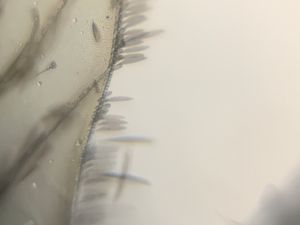
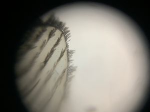
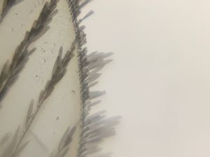
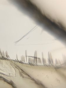
Here are some pictures of the wings. They turned out quite clear even with a good bit of digital zoom.
The last sample was totally unexpected! I was reviewing and deleting the photos I took, when my mom swatted a mosquito above my head. Into the Foldscope it went.
Here are some pictures of the wings. They turned out quite clear even with a good bit of digital zoom.
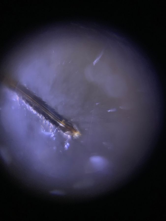
Then I came to the proboscis; the part which the mosquito uses to feed on blood.
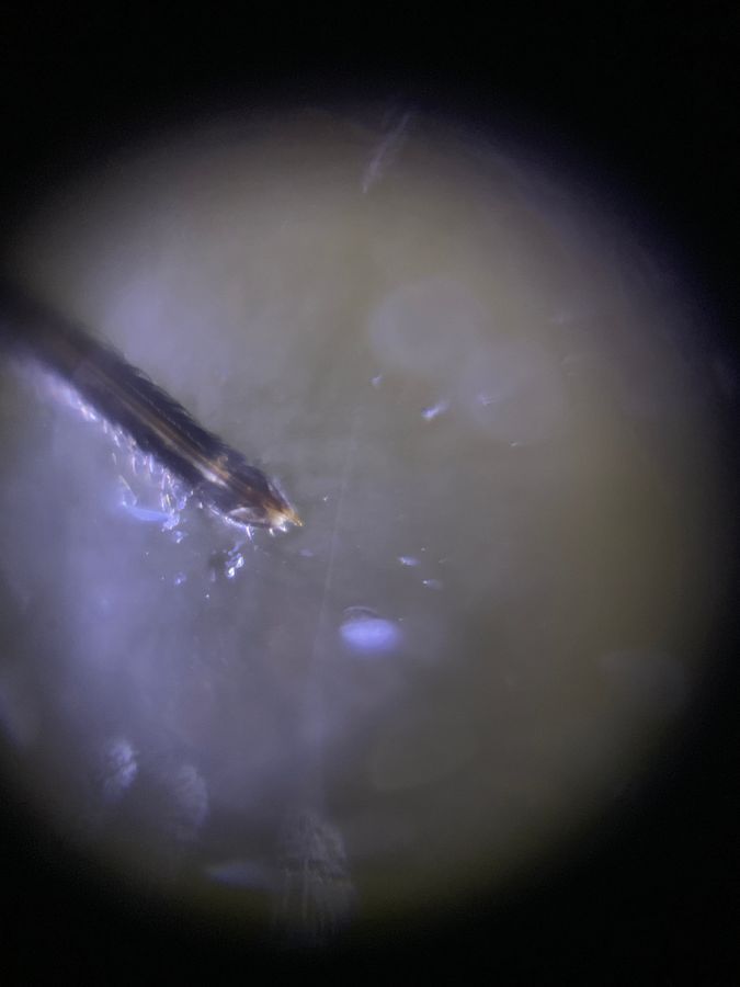
View in Media Gallery
There is a smaller tube inside the main proboscis, which it uses to inject saliva that numbs your skin. Then it sucks blood through the larger tube.
View in Media Gallery
CookerBird #EarthDay
Sign in to commentNobody has commented yet... Share your thoughts with the author and start the discussion!
 0 Applause
 0 Comments