Outer and inner onion epidermises – stained at home!
 Apr 28, 2021 • 10:56 AM UTC
 Unknown Location
 140x Magnification
 Microorganisms
Diya A
Learn about the author...
13posts
9comments
1locations
View in Media Gallery
The first time I sampled an onion peel was a bit different from the introductory experiment in a biology textbook (I observed the outer epidermis, without staining it). Till now I haven’t been able to get the same “3D” effect again (https://microcosmos.foldscope.com/?p=197449), but today’s observations were equally beautiful!
This time, I observed and compared the outer and inner epidermises of an onion separately, and stained them with a homemade food colour stain too!
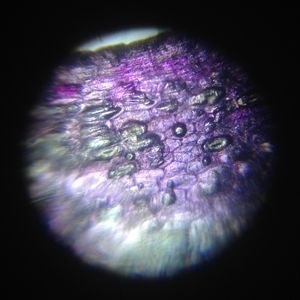
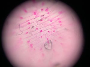
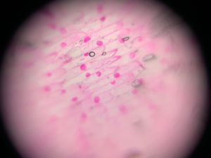
Outer Epidermis (convex side of onion layer)
This is the thin purplish skin on the layers of an onion. It doesn’t peel off in a large, continuous piece.
This time, I observed and compared the outer and inner epidermises of an onion separately, and stained them with a homemade food colour stain too!
Outer Epidermis (convex side of onion layer)
This is the thin purplish skin on the layers of an onion. It doesn’t peel off in a large, continuous piece.
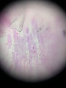

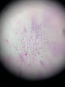
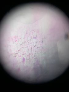
The cell nuclei are comparatively small. The cell walls are thinner and the cytoplasm takes up less stain.
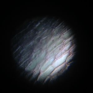
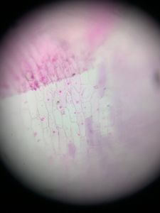
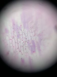
Inner Epidermis (concave side of onion layer)
This membrane is translucent to transparent. It peels off easily in a single piece. The inner epidermis (stained with safranin) is viewed as a first introduction to cells in school.
Inner Epidermis (concave side of onion layer)
This membrane is translucent to transparent. It peels off easily in a single piece. The inner epidermis (stained with safranin) is viewed as a first introduction to cells in school.
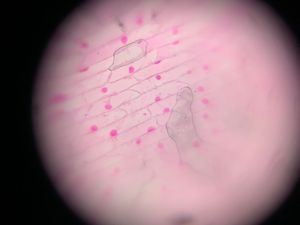
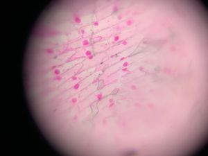
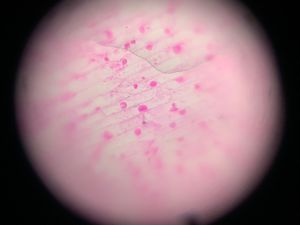
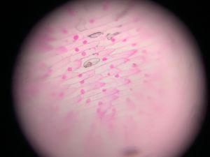

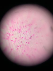
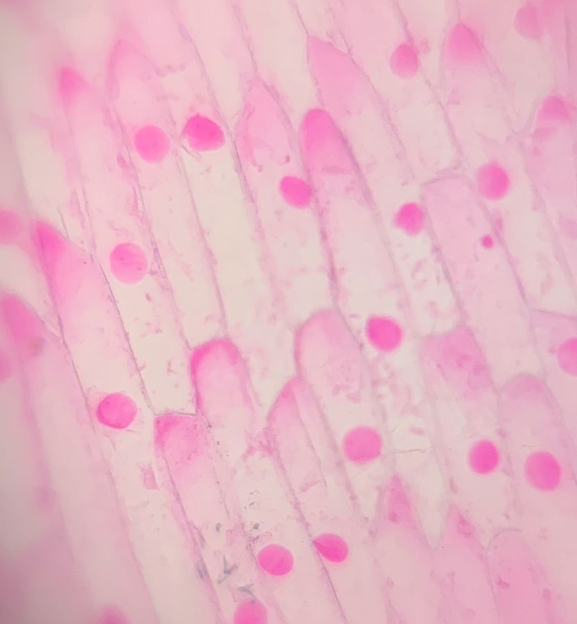
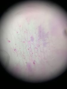
The nuclei of these cells are huge! The cytoplasm is more deeply stained, and cell walls are more distinct.
It is a mystery why these cells – especially the nuclei – are so different in structure! Both are epidermal, and carry out the same function. And they are less than half a centimetre apart, separated by a single layer of onion flesh!
CookerBird
It is a mystery why these cells – especially the nuclei – are so different in structure! Both are epidermal, and carry out the same function. And they are less than half a centimetre apart, separated by a single layer of onion flesh!
CookerBird
Sign in to commentNobody has commented yet... Share your thoughts with the author and start the discussion!

 0 Applause
 0 Comments