Roasted seaweed snack
 May 23, 2021 • 3:34 PM UTC
 Unknown Location
 140x Magnification
 Microorganisms
Diego Huyke
Learn about the author...
1posts
0comments
1locations
View in Media Gallery
Presented are the methods and experimentally obtained images of a roasted seaweed snack under magnification via a Foldscope. This exercise was performed as part of Stanford’s BIOE 301C: Diagnostics Devices Lab.
Methods . A Foldscope was obtained and assembled using the typical procedure. To hold the sample, the paper slides with rectangular windows ( Fig. 1 ) was used. The seaweed used for experiments was Trader Joe’s Wasabi Roasted Seaweed Snack .
Methods . A Foldscope was obtained and assembled using the typical procedure. To hold the sample, the paper slides with rectangular windows ( Fig. 1 ) was used. The seaweed used for experiments was Trader Joe’s Wasabi Roasted Seaweed Snack .
View in Media Gallery
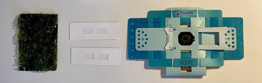
Fig. 1 . Left to right are the roasted seaweed snack, paper Folscope slides, and assembled Foldscope. To mount the seaweed sample, a small (~ 2 x 2 mm) strip of seaweed was placed on a clear adhesive strip. The strip itself was bonded to a paper slide ( Fig. 2 ). Finally, an additional paper slide (used as a spacer) was placed on top of the original slide and mounted on the Foldscope ( Fig. 3 ).
View in Media Gallery
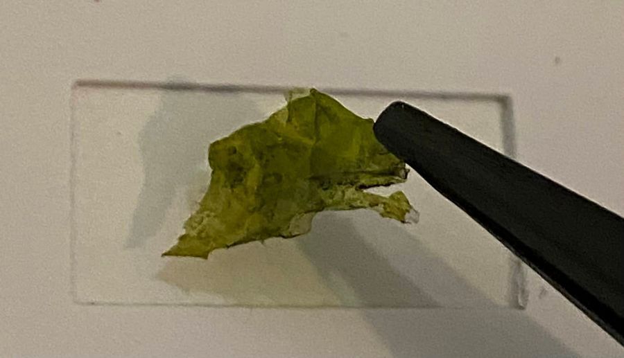
Fig. 2 . Seaweed sample placement on clear adhesive slide.
View in Media Gallery
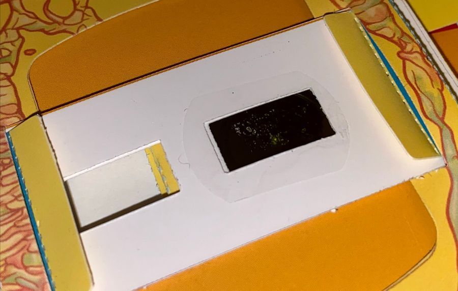
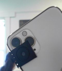
Fig. 3 . Paper slide mounted on Foldscope. To obtain images, the magnetic disk was bonded (via double sided adhesive) to the telephoto lens of an iPhone 11 Pro ( Fig. 4 ). The telephoto lens allows greater zoom-in into the field-of-view enabled by the Foldscope. Hence, better images can be obtained. One challenge, however, is that the default iPhone camera software does not allow selection of this specific lens. The Ullman Indirect app allows selection of the telephoto lens as well as some other convenient features for free.
View in Media Gallery
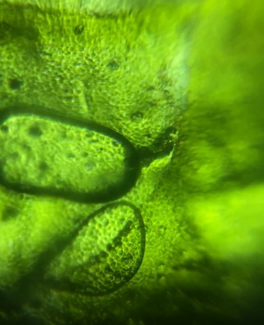
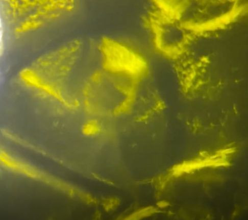
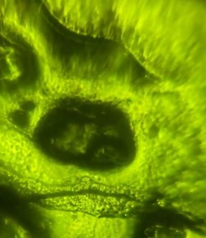
Fig. 4. iPhone 11 Pro with magnetic disk bonded to telephoto lens. Results . The resulting images show, with surprising resolution the components of the seaweed. Most distinctively, we can see the presence of sharp walls (demarcated by the dark regions) which likely correspond to cell walls ( Figs. 5 and 6 ).
View in Media Gallery
Fig. 5 .
View in Media Gallery
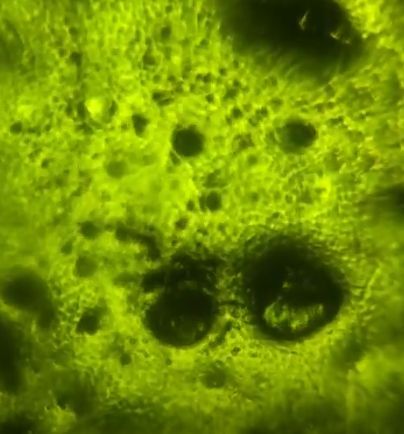
Fig. 6 . Surprisingly, the roasted and salted seaweed appears to retain a lot of cellular complexity when inspected by the Foldscope. This indicates that the seaweed cells are tough and at least somewhat resisting to the drying and roasting process. More images are shown below ( Figs. 7 and 8 ). The dark spots on the seaweed may be attributable to flakes of salt.
View in Media Gallery
Fig. 7 .
View in Media Gallery
Fig. 8 .
Sign in to commentNobody has commented yet... Share your thoughts with the author and start the discussion!
More Posts from Diego Huyke
No more posts from this author.