Observing Micro-Bubbles
 May 24, 2021 • 4:50 PM UTC
 Unknown Location
 140x Magnification
 Unknown
Ziv Lautman
Learn about the author...
1posts
0comments
1locations
View in Media Gallery
I was curious to see if the Lipid Shell Micro-Bubbles we’ve been using in our lab as a contrast agent for Optical Coherence Tomography will be visible with the Foldscope.
The Micro-Bubbles I examined had a C3F8 gas core, wrapped in a lipid shell, and their diameter ranged from 0.6 micron to 1.2 micron. Approximately 5 microliter sample with an unknown concentration of micro-bubbles was loaded on a glass slide.
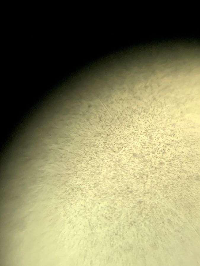
The image below shows the full field of view, as seen with the Foldscope. the micro-bubbles are indeed visible, mainly at the center of the image which is in focus.
The Micro-Bubbles I examined had a C3F8 gas core, wrapped in a lipid shell, and their diameter ranged from 0.6 micron to 1.2 micron. Approximately 5 microliter sample with an unknown concentration of micro-bubbles was loaded on a glass slide.
The image below shows the full field of view, as seen with the Foldscope. the micro-bubbles are indeed visible, mainly at the center of the image which is in focus.
View in Media Gallery
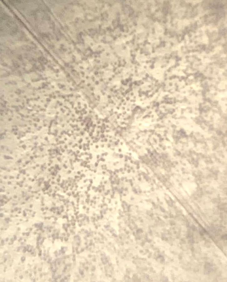
The image below shows the micro-bubbles with maximum zoom (iPhone XR). As can be seen, most of the micro-bubbles are clamped together, since their concentration is high and they have been stored in a vial for the past few months.
View in Media Gallery
Micro-bubbles as seen with a Foldscope Overall, Foldscope is a powerful optical microscope that enables microscopic work anywhere, for only $2. Within a few minutes of easy and fun setup, I was already imaging!
Sign in to commentNobody has commented yet... Share your thoughts with the author and start the discussion!
More Posts from Ziv Lautman
No more posts from this author.