Cells: The Fundamental Units of Life
 May 29, 2021 • 7:44 AM UTC
 Unknown Location
 140x Magnification
 Microorganisms
Thuận Nguyễn Diệu
Learn about the author...
6posts
5comments
1locations
View in Media Gallery
Cell is the unit of life. All living organisms are constructed from cell
View in Media Gallery
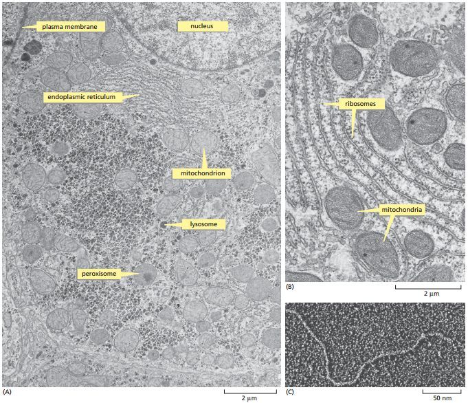
The fine structure of a cell can be seen in a transmission electron microscope.
(A) thin section of a liver cell showing the enormous amount of detail that is visible. Some of the components to be discussed later in the chapter are labeled; they are identifiable by their size and shape
(B) a small region of the cytoplasm at higher magnification. the smallest structures that are clearly visible are the ribosomes, each of which is made of 80–90 or so individual large molecules.
(C) portion of a long, threadlike DNa molecule isolated from a cell and viewed by electron microscopy. (a and B, courtesy of Daniel S. Friend; c, courtesy of Mei Lie Wong.)
View in Media Gallery
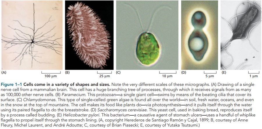
A bacteria cell say a Lactobacillus in a piece of cheese is a few micrometers and it’s about 25 times smaller than the width of a human hair. A frog egg which is also a single cell has a diameter of about 1 millimeter. Figure 1-1 show us that cells vary as widely in their shape, for instance, a typical nerve cell in our brain. A paramecium in a drop of pond water is shaped like a submarine and is covered with thousands of cilia.
View in Media Gallery
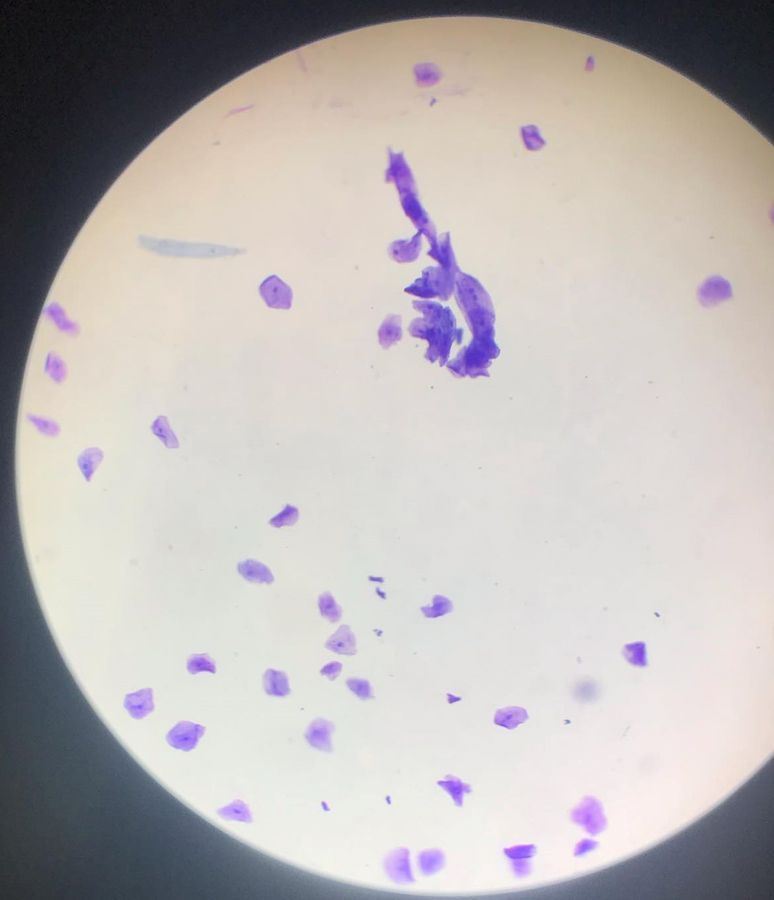
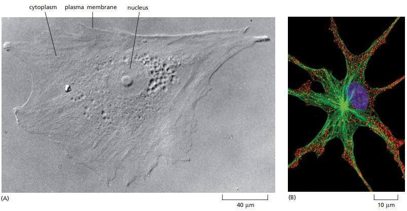
Some of the internal structures of a living cell can be seen with a light microscope
View in Media Gallery
Human cheek cell (EM)
View in Media Gallery
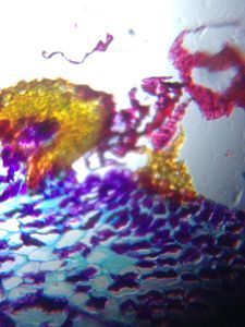
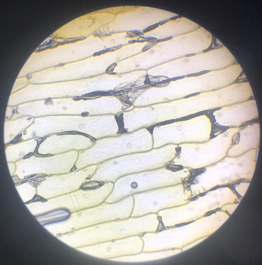
Onion epidermal cell (EM)
View in Media Gallery
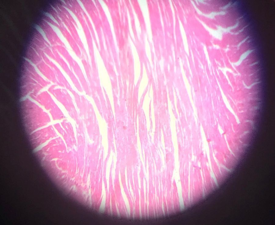
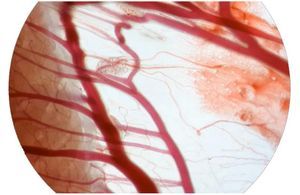
Vertical section of heart (EM)
View in Media Gallery
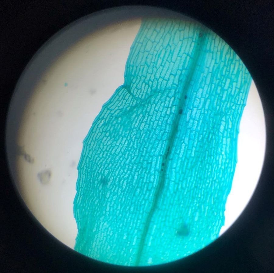
Funaria Hygrometrica (EM)
View in Media Gallery
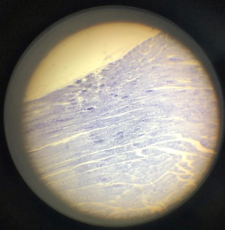
Skeletal muscle (EM)

View in Media Gallery
Vertical section of cardiac muscle tissue (EM) Reference: Essential Cell Biology. 4th Edition 2013; Photos experment when I have learned General Biology Labwork for 1 year.
Sign in to commentNobody has commented yet... Share your thoughts with the author and start the discussion!
 0 Applause
 0 Comments