Imaging a small garden spider
 Aug 04, 2021 • 3:56 AM UTC
 Unknown Location
 140x Magnification
 Unknown
Groovy Greek
Learn about the author...
1posts
0comments
1locations

View in Media Gallery
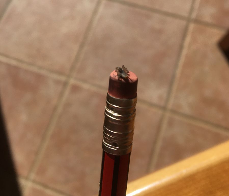
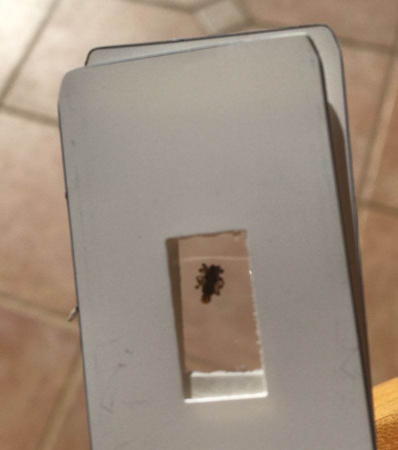
Methods: A small garden spider (species unknown, genus unknown) was sacrificed using the terminal rubber tip of a HB pencil ( Fig. 1 ) at around midday on Saturday 31st July 2021 somewhere in the Western Cape, South Africa. The unprocessed specimen was placed on the plastic Foldoscope slides according to manufacturer’s instructions ( Fig. 2 ). The specimen was then imaged at x140 using natural sunlight, unperturbed from the minimal clouds in the sky. For imaging, the camera lens of an iPhone 6s was magnetically attached (by taping a magnet supplied in the Foldscope classroom kit to the phone) to the blue side of the Foldscope.
View in Media Gallery
Figure 1: Sacrificial apparatus used for the small garden spider.
View in Media Gallery
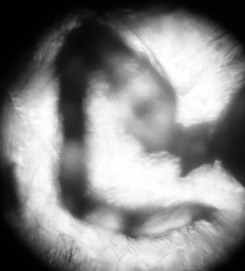
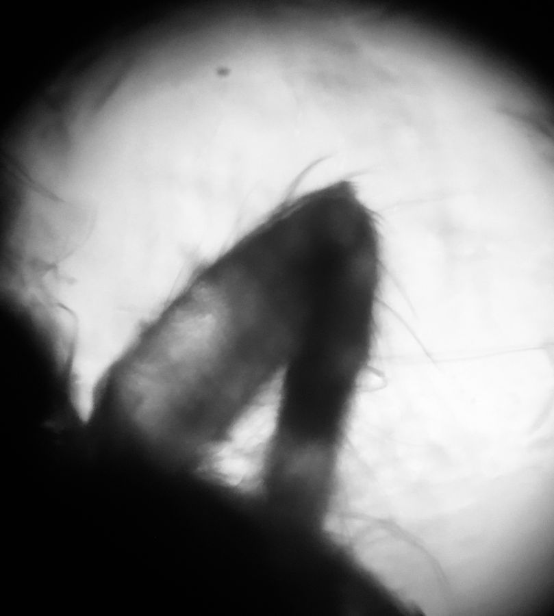
Figure 2: Placement of specimen onto the Foldoscope slides prior to imaging. Results and discussion: Some parts of the spider were more difficult to visualise than others. The limbs were particularly easy to image, and were best observed using a black and white camera filter. The best images, featured below ( Figs. 3 & 4 ), demonstrated visible limb pigmentation which seemed to be aggregated at the limb flexures. A unique feature included in Fig. 3 was the junction between the limb and the body. Higher resolution imaging would be required to better discern the structure of this junctional unit. Hairs appeared to be uniformly and widely distributed. It must be noted though that hairs appeared to be particularly well-concentrated at the tip of limb shown in Fig. 3 . Whilst was difficult to discern the hair morphology from Fig. 3 , the better image focus offered in Fig. 4 showed some variation in hair length. The longer hairs observed around the limb flexure and on the body ( Fig. 4 ), coupled with the higher concentration observed at the terminus of the limb shown in Fig. 3, perhaps suggest that the hairs are a sensory adaptation for the spider to best acclimatise to its changing environments. Future studies could explore the relationship between the hair distribution, length and possible specific functions to guide the spider’s survival in the (sometimes) hostile garden environment.
View in Media Gallery
Figure 3: Spider leg was visibly pigmented at the limb flexures. The junction between the limb and body was visible. Hairs were uniformly distributed with similar lengths throughout. Magnification: x140.
View in Media Gallery
Figure 4: A less contrasted image of another limb. Hairs appear to be more in-focus than in Fig. 3, and also appear to be of variable length. Magnification: x140. Conclusion: This investigation was a fun way to get closer to creatures that scare me, and an opportunity to immerse myself in some of the simpler observational joys of life.
Sign in to commentNobody has commented yet... Share your thoughts with the author and start the discussion!
More Posts from Groovy Greek
No more posts from this author.