My Dictyostelium story
 Oct 23, 2021 • 7:01 PM UTC
 Unknown Location
 140x Magnification
 Microorganisms
Laks Iyer
Human observer of life. https://sukshmadarshin.wordpress.com
97posts
1255comments
5locations

View in Media Gallery
It was some 33 years almost to the date. There were three of us, partners in science; Srinivas V., Vinay T, and myself. We had just read this wonderful book on slime molds by William Loomis and we simply had to see this form of life. The Ruia college microbiology department is an unusual one. They promoted the pursuit of research problems from the first day of our undergrad. Students wrote a research plan and the department provided the reagents and other facilities after perusing the proposal. We would usually do our experiments after college hours. It was an informal program and the three of us (and many like us before and after) still fondly recollect our first steps in the sciences in the department and those wonderful teachers.
So we wrote to the authority of Dictyostelium in India, Dr. Vidyanand Nanjundiah, and to our surprise, he sent us Dictyostelium cultures along with its food, a bacterium, probably E. coli . Dictyostelium is what is informally called a slime mold. It isn’t a fungus but belongs to a group of organisms called the amoebozans (of which Amoeba proteus is a member). Within Amoebozoans, it belongs to the mycetozoans of which the dog vomit-like slime molds are members ( click the numbers to see older foldscope posts on a slime mold of the plasmodial kind, 1 and 2 ). There are two kinds of mycetozoans, the cellular slime molds e.g. Dictyostelium and the plasmodial slime mold such as Physarum . Dictyostelium has a remarkable life cycle. Normally these are solo amoebae that under starvation conditions, aggregate together and form a slug. This aggregation is due to a soluble cyclic nucleotide signal that permeates the surrounding. The Slug now a multicellular organism moves around to another spot, fixes itself and differentiates into a fruiting body (this looks like a fungal fruiting body). I initially thought they forage as a slug, but it seems more like they disperse away from an area where the food resources are low.
You can imagine how excited we were when we received the cultures. We set up the experiment, pouring the bacterium on an agar plate and Dictyostelium on it. In a couple of days, we saw tracks on the plate and something that we thought was a slug. There were high fives and cheers all around until the slug differentiated further into a fruit fly and flew away. Maggots are a problem in tropical climes. I remember we were crestfallen and didn’t have the courage to write to the authority for more samples. We had many experimental successes in our explorations together, but this one wasn’t one.
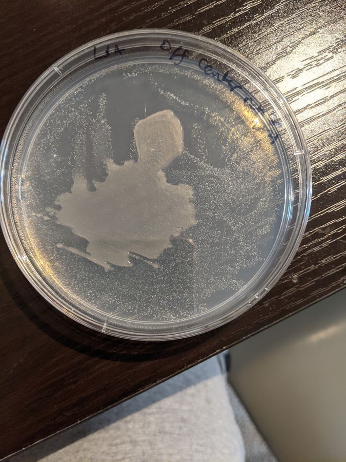
Fast forward and last week, remembering this life-long failure I decided to take this on again. I obtained a commercial culture of Dictyostelium from Carolina biologicals and the bacteria that it feeds on ( E. coli ). I got all the required media and solutions ready and autoclaved them in a dilapidated pressure cooker when everyone was asleep, poured petri plates in the middle of the night to avoid being watched, and inoculated the plates with the bacterium and a solution of Dictyostelium spores (I shall provide detailed protocols in a later post). 24 hours passed and nothing seemed to happen. 48 hours later and this is what I saw. I had dropped a blotch of Dictyostelium onto the center of the plate and something seemed to have grown. The central blotch was thicker than the underlying bacterial colonies.
So we wrote to the authority of Dictyostelium in India, Dr. Vidyanand Nanjundiah, and to our surprise, he sent us Dictyostelium cultures along with its food, a bacterium, probably E. coli . Dictyostelium is what is informally called a slime mold. It isn’t a fungus but belongs to a group of organisms called the amoebozans (of which Amoeba proteus is a member). Within Amoebozoans, it belongs to the mycetozoans of which the dog vomit-like slime molds are members ( click the numbers to see older foldscope posts on a slime mold of the plasmodial kind, 1 and 2 ). There are two kinds of mycetozoans, the cellular slime molds e.g. Dictyostelium and the plasmodial slime mold such as Physarum . Dictyostelium has a remarkable life cycle. Normally these are solo amoebae that under starvation conditions, aggregate together and form a slug. This aggregation is due to a soluble cyclic nucleotide signal that permeates the surrounding. The Slug now a multicellular organism moves around to another spot, fixes itself and differentiates into a fruiting body (this looks like a fungal fruiting body). I initially thought they forage as a slug, but it seems more like they disperse away from an area where the food resources are low.
You can imagine how excited we were when we received the cultures. We set up the experiment, pouring the bacterium on an agar plate and Dictyostelium on it. In a couple of days, we saw tracks on the plate and something that we thought was a slug. There were high fives and cheers all around until the slug differentiated further into a fruit fly and flew away. Maggots are a problem in tropical climes. I remember we were crestfallen and didn’t have the courage to write to the authority for more samples. We had many experimental successes in our explorations together, but this one wasn’t one.
Fast forward and last week, remembering this life-long failure I decided to take this on again. I obtained a commercial culture of Dictyostelium from Carolina biologicals and the bacteria that it feeds on ( E. coli ). I got all the required media and solutions ready and autoclaved them in a dilapidated pressure cooker when everyone was asleep, poured petri plates in the middle of the night to avoid being watched, and inoculated the plates with the bacterium and a solution of Dictyostelium spores (I shall provide detailed protocols in a later post). 24 hours passed and nothing seemed to happen. 48 hours later and this is what I saw. I had dropped a blotch of Dictyostelium onto the center of the plate and something seemed to have grown. The central blotch was thicker than the underlying bacterial colonies.
View in Media Gallery
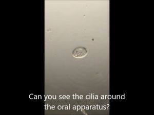
I scooped out a bit of the agar where I had spotted the Dictyostelium and put it on a slide (See main collage) . To my absolute delight, under the foldscope I saw amoebae foraging in the medium. Below is a timelapse. They moved slowly and so I decided to study them using timelapse taking a picture every 10 seconds for about an hour. I have many hours of time-lapse movies, almost as vengeance for that failure 3 decades ago.
I wanted to image the whole petridish to look for any macrophenomena and to my utter delight, I observed the aggregation and the grex stages of Dictyostelium . However, these are low-power scans with my other microscope. I haven’t reproduced this under the foldscope, but do have a plan as to how I can do this. Until then, I just wanted to put the below in the post for completion. These are time-lapses too and I’ll let you enjoy the movies. I am still waiting for the fruiting body to form. I can sleep well tonight and yes I did tell my dear friends about my success and they can sleep well too. 🙂
Sign in to commentNobody has commented yet... Share your thoughts with the author and start the discussion!

 0 Applause
 0 Comments
_300x300.jpeg)