Blister-y Fun
 Nov 24, 2021 • 11:33 AM UTC
 Unknown Location
 140x Magnification
 Unknown
Catherine Keim
Learn about the author...
1posts
0comments
1locations

View in Media Gallery
This past weekend, I had the fortune of cheering my big
sister on as she ran in her very first marathon in Philadelphia! One of the
consequences of training for long distance running, however, is that my sister’s
feet are…gnarly. As a former dancer, I’m used to feet covered in bruises and
blisters, but I will spare everyone a picture of my sister’s runner’s feet.
View in Media Gallery
My sister (orange shirt) and her friends post-marathon in front of the Philadelphia Art Museum One of my sister’s blisters interested us a lot, because the
outer layer of the skin had become detached at certain parts, and there was another blister developing underneath (it sounds a lot worse than it is, but I will
once again spare everyone a picture). With the excitement of knowing that we
could use my Foldscope to look at her skin, we decided to cut off a small
sliver of the detached portion of the blister and make a slide out of it. What
we saw was super interesting, with what appears to be cell walls and different
thicknesses of the skin. This led me to do some more research about what
blisters actually are and how they are formed.
View in Media Gallery
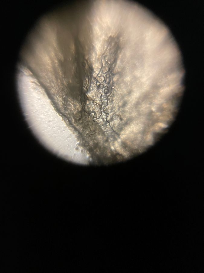
A closer look at the skin off my sister’s blister
View in Media Gallery
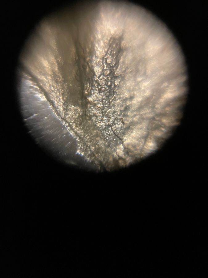
A closer look at the skin off my sister’s blister
View in Media Gallery
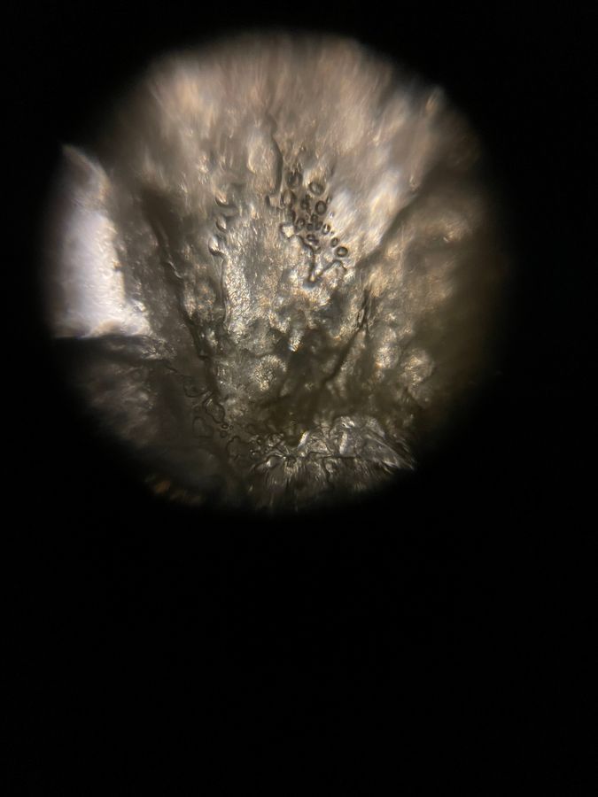
A closer look at the skin off my sister’s blister According to Johns Hopkins Medicine, blisters are bubbles of skin that contain fluid. They can be caused by allergic reactions, infections, and injury like friction or burns. Additionally, blisters form in the upper layer of the skin, the epidermis, to protect the layers of skin below (Medical News Today). In the case of my sister’s blisters, which formed most likely from friction, these blisters come from fluid filling gaps in the epidermis that are created when the tissue breaks down by repeated rubbing. This fluid supports tissue growth, and it slowly disappears as skin grows beneath the blister.
Because my sister’s blister was beginning to fall off, we can assume that her skin was healing. However, it looked like there was another blister forming beneath, and I hypothesize that this was the formation of a callus! Calluses are formed when repeated pressure causes skin to die and form a protective surface (University of Michigan Health). While the Foldscope gave me insight into what was actually going on in the outer layer of my sister’s skin, I would love to be able to look at the layers that are being formed in her new callus (but alas, that would require us to open up her toe, and we don’t have the tools for that). The images from the Foldscope on her current sample allowed me to look at the small folds in her blister’s layers, so at least there is that.
While this experience has not made me want to become a dermatologist, I’m still very excited to see how my sister’s feet heal and what they look like as they protect themselves against long distance running.
I conducted this project as part of Professor Pringle’s EEB321 class at Princeton University.
References:
https://www.hopkinsmedicine.org/health/conditions-and-diseases/blisters
https://www.medicalnewstoday.com/articles/264783#causes
https://www.uofmhealth.org/health-library/ug2399
Sign in to commentNobody has commented yet... Share your thoughts with the author and start the discussion!
More Posts from Catherine Keim
No more posts from this author.