Foldscope day at the London School of Hygiene and Tropical Medicine: specimen pictures
 Dec 05, 2016 • 10:50 AM UTC
 Unknown Location
 140x Magnification
 Microorganisms
Ailie Robinson
Learn about the author...
1posts
0comments
1locations
View in Media Gallery
In November we ran a ‘disease diagnostics’ session here in the teaching lab at LSHTM, and local school students came in to learn about diagnostics and view specimens – via Foldscopes! We discussed malaria and the Oriental liver fluke ( Chlonorchis sinensis ), then the students folded their Foldscopes and looked at the sections: we had malaria-infected brain ( Plasmodium falciparum ), with signs of micro-hemorrhaging and parasite pigment, as well as the liver sections. These are some of the pictures taken!
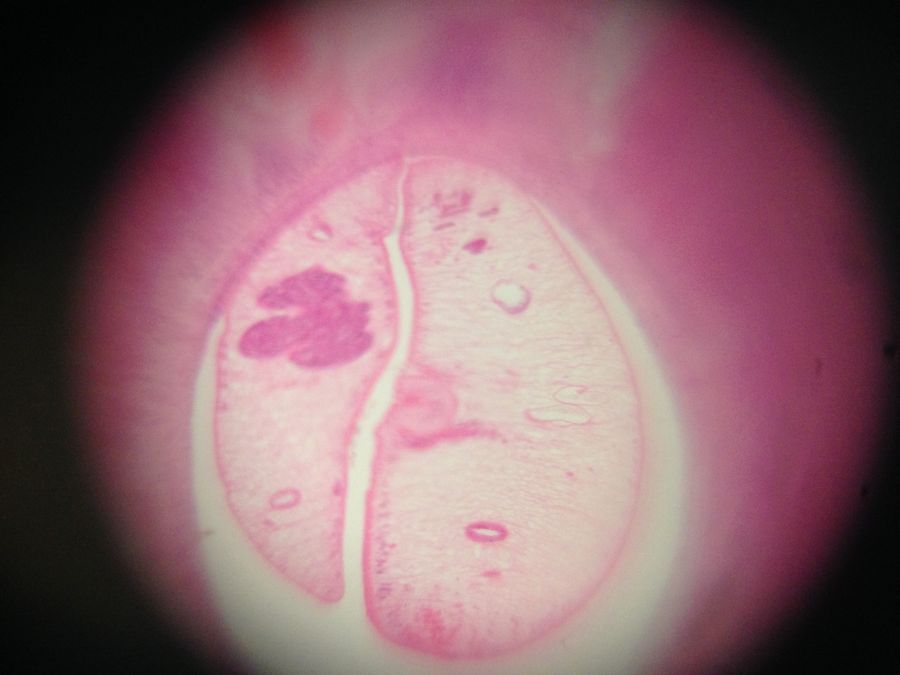
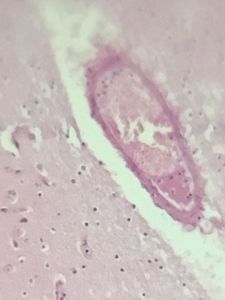
Foldscope number: 0001CFC71A56 , liver fluke: the two small circles on either side (that look like eyes) are the two intestinal branches (diagnostic for Chlonorchis sinensis ). The fluke is in a bile duct, this may be two flukes or one that is folded in two. The bile duct around it is enlarged and with inflammatory cells.
Foldscope number: 0001CFC71A56 , liver fluke: the two small circles on either side (that look like eyes) are the two intestinal branches (diagnostic for Chlonorchis sinensis ). The fluke is in a bile duct, this may be two flukes or one that is folded in two. The bile duct around it is enlarged and with inflammatory cells.
View in Media Gallery
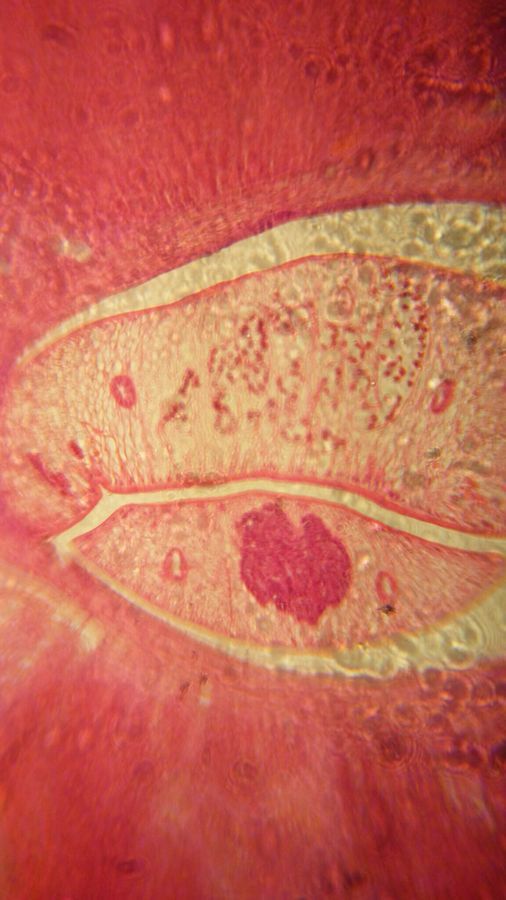
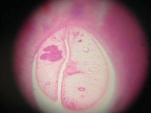
Foldscope number: 0001 493B B5C5 , close-up of detail on a liver fluke, then two brain images; the first showing hemorrhaging (red areas in the amorphous pink tissue [brain]), the second focused on a blood vessel, with dark malaria pigment inside the vessel. This is caused by schizonts – one of the stages of Plasmodium’s asexual replication – lodging in the vessels and causing blockages, leading to the characteristic pathology of cerebral malaria. The dark pigment is a waste product of the parasites, and is derived from haemoglobin.
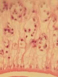
Foldscope number: 0001 576CAAEF , liver fluke as above, with zoom in on area of liver section with many inflammatory cells.
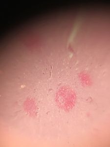
Foldscope number: 0001 9BEA 448C, malaria brain: haemorrhaging and pigment clearly visible in pinkish area in the centre
View in Media Gallery
More photos from the day to follow!
Sign in to commentNobody has commented yet... Share your thoughts with the author and start the discussion!
More Posts from Ailie Robinson
No more posts from this author.