Exploring the Deep Blue Sea
 Dec 13, 2016 • 7:41 PM UTC
 Unknown Location
 140x Magnification
 Microorganisms
Julie Fogarty
Learn about the author...
1posts
2comments
1locations
View in Media Gallery
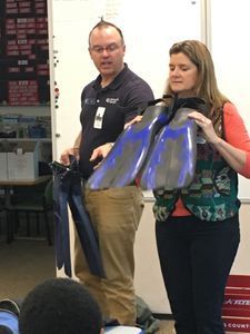
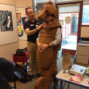
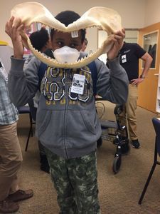
Sometimes an awesome opportunity presents itself to you. That’s what happened to our class at the Lucile Packard Children’s Hospital School. Dan A., one of the shark divers from the Monterey Bay Aquarium, got connected with one of our teachers (Kathy H.) about bringing an aquarium-like learning experience to our hospital school students. He brought shark jaws and teeth, talked about diving gear, and discussed what it is like to work at the aquarium.
Dan also wanted to bring us some plankton samples to look at under microscopes so that kids could get an idea of what lives in ocean water. Sounds awesome, right? The problem is that we are a small school with just two classrooms, and we don’t have a class microscope. At first, we were disappointed that our kids wouldn’t get to look at these amazing creatures. Enter the Prakash Lab. One of our graduate student volunteers (Julie F.) and our Stanford faculty sponsor (Andy S.) had both previously mentioned the amazing Foldscope project. Julie reached out to the Prakash Lab, and Manu generously offered a class set of Foldscopes for our students to use to explore the exciting world of plankton.
Here is the first sample we viewed using the Foldscope. It is a copepod – a small, planktonic crustacean found in the sea and nearly all freshwater habitats. We’ll show a much more deliberate video of the copepod later on, but what is exciting about this video is that we were able to see the ciliates (small, oblong shapes) swimming around the copepod. Ciliates serve as food for the copepod and are very small. You’ll hear Dan remark that these ciliates are difficult for them to see, even with the microscopes they have in their lab. We were able to see them on our novice tour with the Foldscope – isn’t that AMAZING?!?
Here is the first sample we viewed using the Foldscope. It is a copepod – a small, planktonic crustacean found in the sea and nearly all freshwater habitats. We’ll show a much more deliberate video of the copepod later on, but what is exciting about this video is that we were able to see the ciliates (small, oblong shapes) swimming around the copepod. Ciliates serve as food for the copepod and are very small. You’ll hear Dan remark that these ciliates are difficult for them to see, even with the microscopes they have in their lab. We were able to see them on our novice tour with the Foldscope – isn’t that AMAZING?!?
View in Media Gallery
Here is a much more deliberate video of a single copepod. You can see the head and antennae, the copepod body with five swimming legs, followed by the copepod’s “butt.”
View in Media Gallery
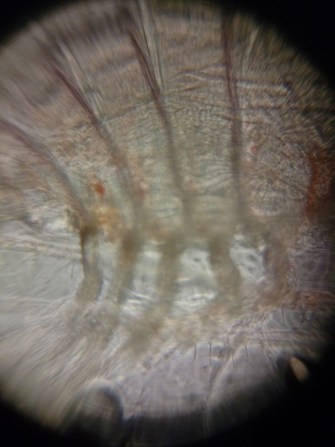
Finally, we looked at a sea urchin in its pluteus larval stage of development. This is the form of the sea urchin that occurs as the embryo changes shape and the skeleton grows. Eventually, a miniature adult sea urchin will hatch and crawl away from the discarded skeleton. The mouth of the sea urchin is opposite the pointed end, amidst the larval arms. The circle you see in the center of the sea urchin pluteus as the camera focuses is the gut, and the long dark lines you see within the body at the same time are skeletal rods. The motion of the sea urchin pluteus is propelled by rows of tiny ciliated cells. Nature is simply BREATHTAKING!
View in Media Gallery
Getting to use the Foldscopes in a class of kindergarteners thru 10 th graders is such a great opportunity. Many of these students haven’t used microscopes in the past, and getting to examine plankton was an experience they won’t soon forget. Thanks, Prakash Lab, for bringing science to life in our hospital classroom!
Sign in to commentNobody has commented yet... Share your thoughts with the author and start the discussion!
More Posts from Julie Fogarty
No more posts from this author.