Alga in my filtered water
 Feb 23, 2015 • 2:21 PM UTC
 Unknown Location
 140x Magnification
 Unknown
Laks Iyer
Human observer of life. https://sukshmadarshin.wordpress.com
97posts
1255comments
5locations
View in Media Gallery
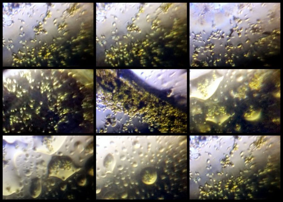
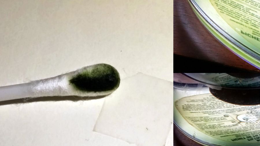
I was about to take a swig from my large flask of filtered water (these Brita flasks have a filtering system as their top), which I keep by my window, when suddenly I noticed a green sheen at the bottom, an unexpected foldscope problem. I drained all the water and scraped the sheen out with a qtip and suspended the green stuff in a drop of water on a slide and covered it with a glass coverslip and taped the sides with cellophane tape.
View in Media Gallery
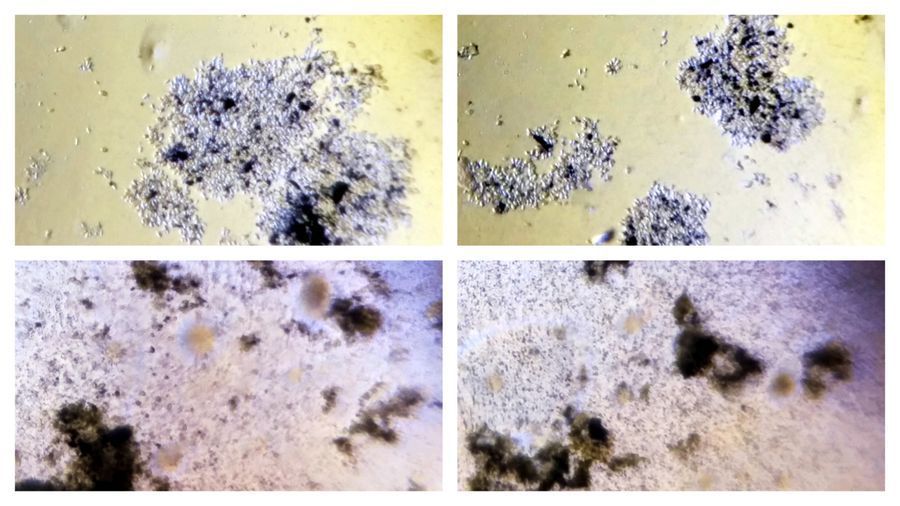
Under the 140x lens, (low power), I noticed tiny oval cells, but the green color was only seen in the clumps.
View in Media Gallery
And so I put it under the high power lens, 400x. It showed a single type of cell, a eukaryotic alga in all likelihood, but I felt frustrated about not seeing the source of the green and so…
View in Media Gallery
I tried and tried to bend and twist the foldscope,since that is often an easy way to align the LED with the condenser if it is mis-aligned. For once the blue light that came through wasnt what I wanted and so I started reading about how the white LED is actually a blue LED with a fluorescent substance that makes it white. Why the inventors of the blue LED got the Nobel prize (and deservedly), all this didnt solve my problem though.
In the meantime, a strange phenomenon emerged, there were bubbles in my field and they were everywhere (about 45 minutes after making the sample). After some juggling, I reassembled the LED and condenser of the foldscope and made a new slide and lo and behold, when the lights were aligned the sight was wonderful…it was a chlorophyte alga no doubt and then it struck me that the alga was photosynthesizing and hence producing oxygen bubbles.
In the meantime, a strange phenomenon emerged, there were bubbles in my field and they were everywhere (about 45 minutes after making the sample). After some juggling, I reassembled the LED and condenser of the foldscope and made a new slide and lo and behold, when the lights were aligned the sight was wonderful…it was a chlorophyte alga no doubt and then it struck me that the alga was photosynthesizing and hence producing oxygen bubbles.
View in Media Gallery
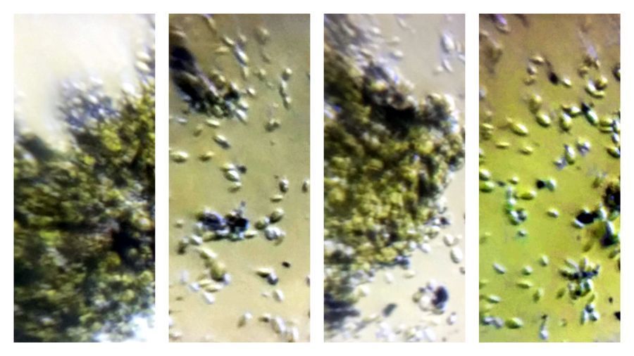
Greedy to see more, I put the sample under an oil-immersion lens of my compound microscope (celestron — 1000x) and was satisfied that the problem was solved. Can anyone identify the species?

View in Media Gallery
These algae are tiny and might have entered my flask and accumulated over time as a mono-layer photosynthesizing by the window light. I know for one that my extra-oxygenated water isnt toxic :), but the flask needs a thorough wash. The exercise reminded me of a lesson I received some two and a half decades ago from my inspiring microbiology teacher on the importance of the condenser system (I hope he is reading this).
Below movie shows a bubble pushing the algae under a foldscope 400x. One can also spot some Brownian motion in the alga.
Below movie shows a bubble pushing the algae under a foldscope 400x. One can also spot some Brownian motion in the alga.
Sign in to commentNobody has commented yet... Share your thoughts with the author and start the discussion!

 0 Applause
 0 Comments
_300x300.jpeg)


