A quick post to share some observations
 Mar 04, 2017 • 1:45 AM UTC
 Unknown Location
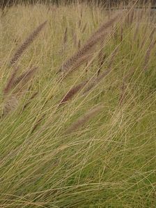
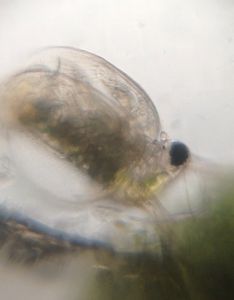
 140x Magnification
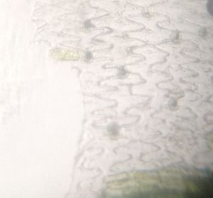
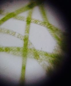
 Microorganisms
Cristina Bosch Esteva
Learn about the author...
19posts
52comments
1locations
View in Media Gallery
Dear foldscope community:
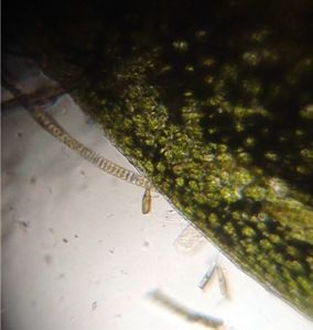
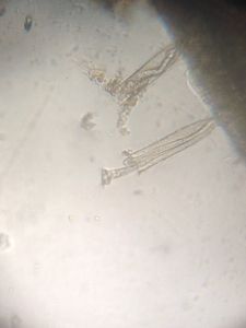
This is going to be a short post due to such a busy time I am having. Just wanted to share with you a bit of the hidden beauty foldscope is allowing many of us to unveal. Isn’t this a gorgeous adventure!
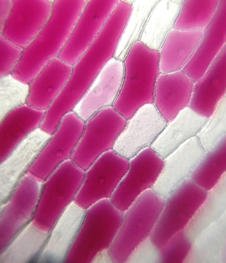
A few days ago I was trying to image epidermal tissue of a red onion in order to register the changes undergone in different osmotic media, that is to say, in water first and in a high concentrated dilution after. As you might know, this is a very common lab experiment accomplished by high school students. To be honest, my goal was to design some easy way to image the process alive, which meant changing from tap water to salty water without moving the slide from the foldscope I was using. I practiced a long cut in a PVC slide y made for this ocassion but it failed, even after three or four tries. Nonetheless, I was completely overjoyed at the images that resulted. Using a foldscope has let me understand we are also playing with light (or light is playing with us) when we observe samples, and this is a fact imposible to facilitate using a regular microscope. Please, watch next images and see what I mean.
This is going to be a short post due to such a busy time I am having. Just wanted to share with you a bit of the hidden beauty foldscope is allowing many of us to unveal. Isn’t this a gorgeous adventure!
A few days ago I was trying to image epidermal tissue of a red onion in order to register the changes undergone in different osmotic media, that is to say, in water first and in a high concentrated dilution after. As you might know, this is a very common lab experiment accomplished by high school students. To be honest, my goal was to design some easy way to image the process alive, which meant changing from tap water to salty water without moving the slide from the foldscope I was using. I practiced a long cut in a PVC slide y made for this ocassion but it failed, even after three or four tries. Nonetheless, I was completely overjoyed at the images that resulted. Using a foldscope has let me understand we are also playing with light (or light is playing with us) when we observe samples, and this is a fact imposible to facilitate using a regular microscope. Please, watch next images and see what I mean.
View in Media Gallery
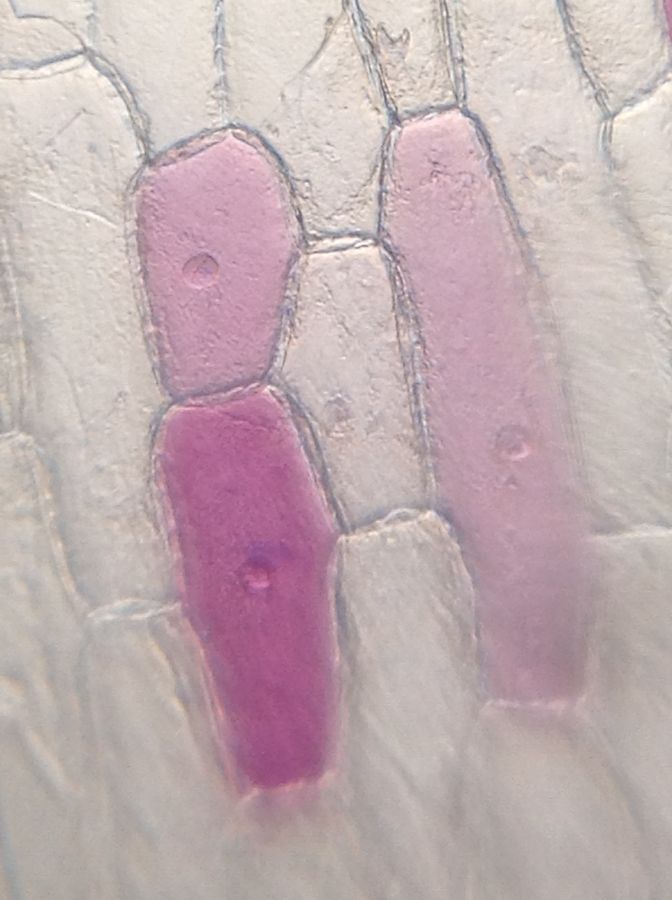
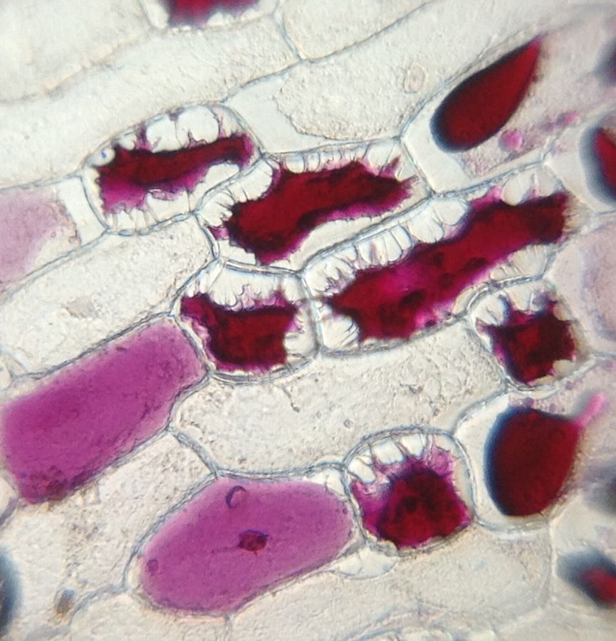
You can almost touch the cells and feel the slight roughness of its surface. I felt amazed!
When the cell underwent plasmolysis, the vacuole containing pigments (anthocyanins, mostly) shrinked and, for the first time in my life, I saw such clear junctions between the cell membrane and the cell wall (Hetchian strands).
When the cell underwent plasmolysis, the vacuole containing pigments (anthocyanins, mostly) shrinked and, for the first time in my life, I saw such clear junctions between the cell membrane and the cell wall (Hetchian strands).
View in Media Gallery
I have gone through these images many, many times and believe they are very beautiful. As I said before, osmosis is a familiar practice for many of us, but foldscope-light duetto has made it different. Simply wanted to let you know.
Till next time
Cristina Bosch
Till next time
Cristina Bosch
Sign in to commentNobody has commented yet... Share your thoughts with the author and start the discussion!
 0 Applause
 0 Comments