Measuring the Foldscope Universe-1: Using the kit scale
 Nov 27, 2017 • 9:52 AM UTC
 Unknown Location
 140x Magnification
 Microorganisms
Laks Iyer
Human observer of life. https://sukshmadarshin.wordpress.com
97posts
1255comments
5locations
View in Media Gallery
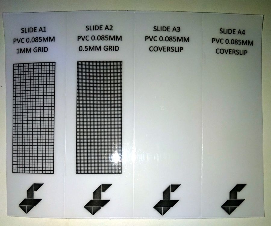
In any scientific endeavor, measurements are a central aspect, and so I was delighted when I saw slide scales in the new foldscope kit. This meant that I could calibrate my phone camera with the slide scales.
View in Media Gallery
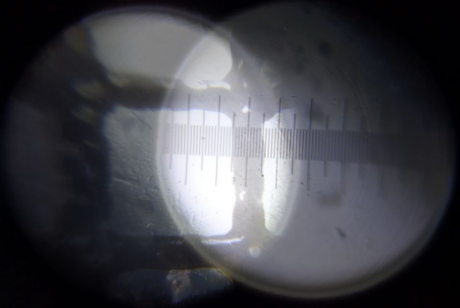
The smallest division on SLIDE A2 is 0.5 mm. Under the foldcsope at no digital zoom, that is quite a distance. It basically spans most of the field. So I decided to find out what the thickness of the line itself is.
View in Media Gallery
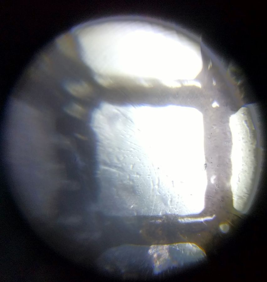
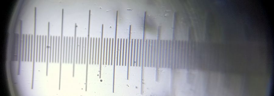
For this, I used a slide scale whose smallest division is 0.01 mm. Thus I obtained, at no digital magnification, the following under the foldscope (140x).
View in Media Gallery
So in essence, I can get the width of each line of the provided slide scales by combining the information from the two images. There are many ways to do this. The simplest way is image merger and that is what I used for the longest period until I discovered ImageJ. Since I have both unprocessed images at the same magnification, I could merge the two to get a superimposed collage. I did this using picasa, but most smart phones have photo collage software that can help you merge the images.
View in Media Gallery
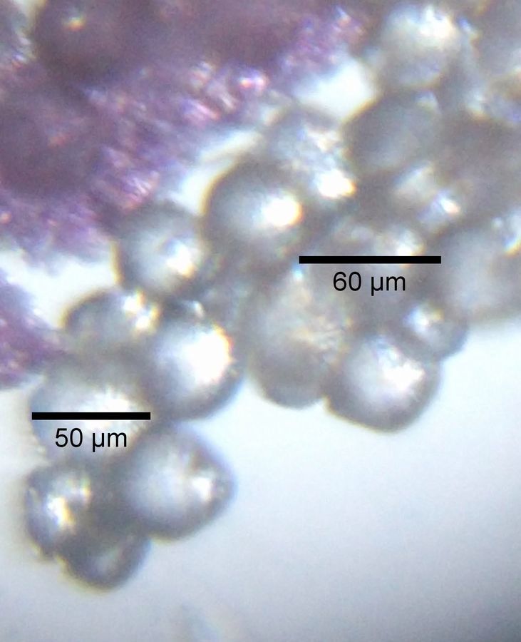
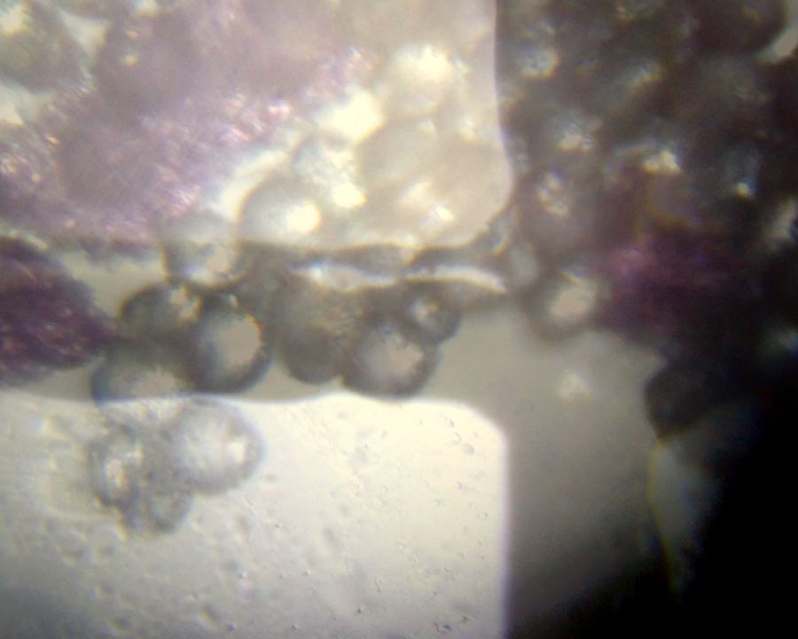
As you can see that each line on the provided slide scale corresponds to 10 divisions of the finer slide scale and hence each line of the slide scale provided in the foldscope kit is about 0.01×10 = 0.1 mm/100 microns thick. So by simply superimposing your image with the image of the slide scale at the same magnification you can get the size of your objects in the image. So for example, here is a (con-scale) merger of the image of christmas cactus pollen and the foldscope kit slide scale. Using my fingers :), I can see that each pollen is a little more than half a line thick, so my best estimate would be about 50-60 microns.
View in Media Gallery
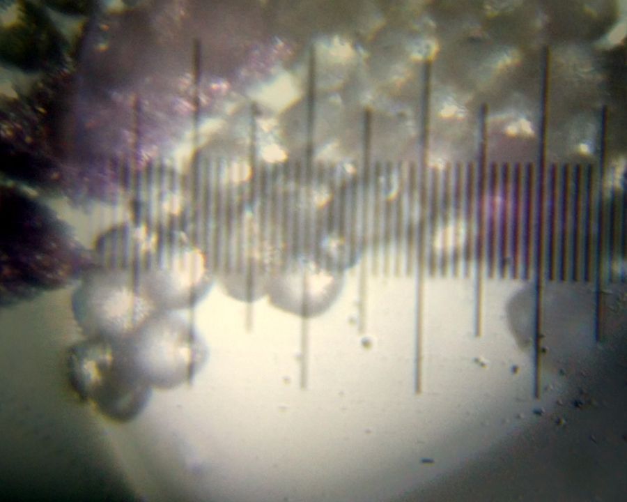
How does that compare to the finer slide scale?
View in Media Gallery
This merger shows that the pollen are about 50-60 microns in size, some longer, some shorter.
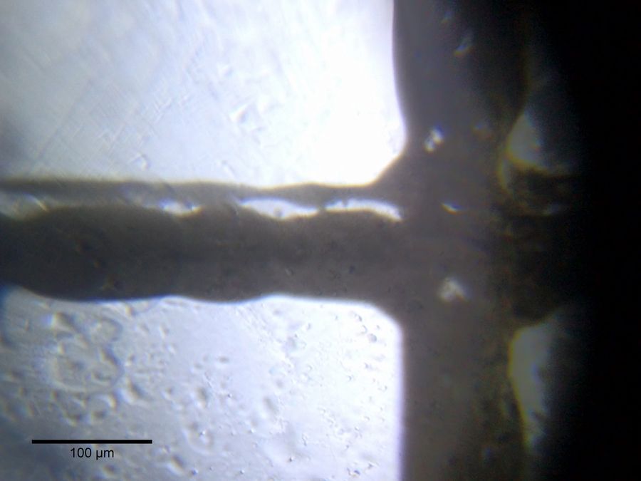
Another useful way is to use a program called ImageJ, a freeware used extensively by microscopists (https://imagej.nih.gov/ij/). Using ImageJ I can measure the number of pixels that corresponds to a certain distance under the foldscope at a certain optical and digital magnification. Once that is fixed, you have a permanent scale for a particular magnification for a particular phone. ImageJ can also compute areas and has other wonderful features. Thus, with ImageJ, I observe that there are 756 pixels corresponding to 100 microns at maximum digital zoom. This information can be used by the software to put a nice looking scale bar at the bottom left.
Another useful way is to use a program called ImageJ, a freeware used extensively by microscopists (https://imagej.nih.gov/ij/). Using ImageJ I can measure the number of pixels that corresponds to a certain distance under the foldscope at a certain optical and digital magnification. Once that is fixed, you have a permanent scale for a particular magnification for a particular phone. ImageJ can also compute areas and has other wonderful features. Thus, with ImageJ, I observe that there are 756 pixels corresponding to 100 microns at maximum digital zoom. This information can be used by the software to put a nice looking scale bar at the bottom left.
View in Media Gallery
Based on this calibration, my pollen are about 50-60 microns. ImageJ can do a lot more and I encourage you to explore it.
View in Media Gallery
One thing to remember is that no measurement is entirely accurate, there is always a bit of variation and error. Finer the scale, lower is your error but variations are common in biological systems. Thus, most measurements are within some kind of a normal distribution. I wish I had learnt that in school and I wish all teachers emphasize this aspect and not punish their students if their acceleration due to gravity value is not exactly 9.8 m/s2 (old wounds still hurt). Here is your chance to experience this aspect of measurement in person.
In the next part, I want to discuss some aspects of scale that interest me in particular– you might call it my philosophy of scale. For now, I hope you could calibrate your scales similarly and make it a point to measure everything in the foldscope universe. You will be surprised as to what you can learn with this.
P.S. I know I can get sharper images and that has to do with my lighting. I am working hard on this.
Sign in to commentNobody has commented yet... Share your thoughts with the author and start the discussion!

 0 Applause
 0 Comments
_300x300.jpeg)


