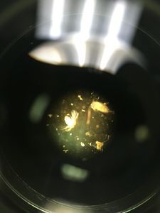
Wet-mount preparation of red alga from Monterey Bay
 Apr 25, 2017 • 1:24 PM UTC
 Unknown Location
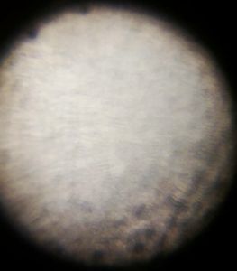
 140x Magnification
 Fungi
Fungi
Jessica Bray
Learn about the author...
6posts
0comments
3locations

View in Media Gallery
I love to I.D. algae in the intertidal, but red algae can be especially cryptic. Here I am preparing my sample with a glass slide, and a glass slip that I am holding together with some Scotch tape so that I can wiggle it into place within my foldscope.
View in Media Gallery
The water in my wet mount is adhering to the algae, and is visible as a little halo. I’m really impressed with the detail I am getting, but since I can see down to the cellular level now, I think I will skip the water next time. That way I can try to examine the number of cells across the width of each branch.

View in Media Gallery
Under the microscope it’s looking consistently like a Polysiphonia! A common branching red alga, that should be found locally. I can’t I.D. this down to the species level, but I can clearly see the pericentral cells as the red globular units in my branches. The water is still visible as a clear border, next time I might dab my algae on a paper towel first.

View in Media Gallery
This was a really easy slide to prepare, which I did by pinching the tip of a filamentous algae off, from a larger piece within a water sample taken from the Coast Guard Jetty again in Monterey. If I were to thin out this very 3-D apical tuft, I just might get lucky enough to see a reproductive body!
Sign in to commentNobody has commented yet... Share your thoughts with the author and start the discussion!

 0 Applause
 0 Comments