Patterns, and more patterns
 May 14, 2017 • 9:40 PM UTC
 United States
 140x Magnification
 Plants
Shannon Myers
Learn about the author...
3posts
0comments
2locations

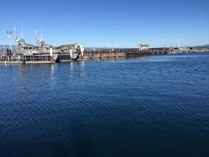
Hi there, this is Shannon again returning with my second Foldscope post. On a drizzly, grey day I set out to explore a gorgeous rocky beach north of Fort Bragg, CA. My walk out to the coastal bluffs revealed a magnificent expanse of rocky intertidal beaches. In the distance, a group of sea lions watched carefully from a stone perch and eventually slid back into the safety of the ocean.


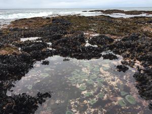
After descending a steep traverse, I faced an upper intertidal zone with pool after pool of amazing intertidal life. The brightly-colored anemones, sea stars, and iridescent algae and mussel shells transformed a dreary Northern California day on the verge of spring into a wondrous adventure.
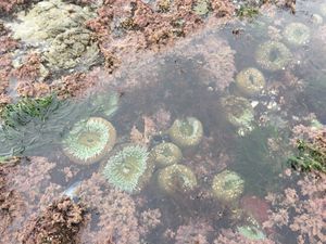
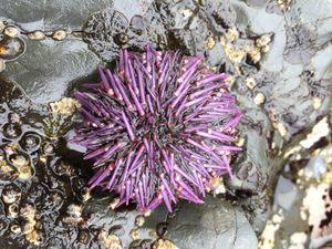

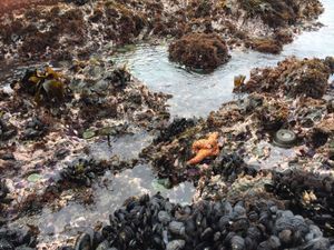
A few species that caught my attention included: California mussel ( Mytilus californianus ), giant green anemone ( anthopleura xanthogrammica ), surfgrass ( Phyllospadix ), purple sea star ( Pisaster ochraceus ), purple sea urchin ( Strongylocentrotus purpuratus ), and a variety of macroalgae.
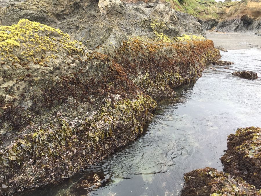
Every time I visit a rocky intertidal, I am always struck by the zonation pattern of macroalgae, or seaweed. Empowered to investigate physical characteristics too small to see, I collected samples of three seaweeds that displayed a distinct vertical zonation pattern with the hope that the Foldscope may shed some light on this subject. The bright yellowish-green new-growth Porphyra perforata dominated the top zone, followed by reddish-brown Mastocarpus papillatus in the mid-zone, and lastly by Mazzaella flaccida along the lower vertical range nearly touching the low-tide pools. Generally it is thought that physical factors (temperature, desiccation, etc.) set the tolerance limits in the upper intertidal, and that biological factors (primarily predation) set the lower limits for many species of both invertebrate and seaweed species. My question was whether the Foldscope would allow me to see any cellular differences between the three species of seaweed that so clearly occupied different intertidal positions. Zonation patterns assuredly are driven by a combination of physical and chemical characteristics, but my equipment limited me to investigating only the physical differences.
Every time I visit a rocky intertidal, I am always struck by the zonation pattern of macroalgae, or seaweed. Empowered to investigate physical characteristics too small to see, I collected samples of three seaweeds that displayed a distinct vertical zonation pattern with the hope that the Foldscope may shed some light on this subject. The bright yellowish-green new-growth Porphyra perforata dominated the top zone, followed by reddish-brown Mastocarpus papillatus in the mid-zone, and lastly by Mazzaella flaccida along the lower vertical range nearly touching the low-tide pools. Generally it is thought that physical factors (temperature, desiccation, etc.) set the tolerance limits in the upper intertidal, and that biological factors (primarily predation) set the lower limits for many species of both invertebrate and seaweed species. My question was whether the Foldscope would allow me to see any cellular differences between the three species of seaweed that so clearly occupied different intertidal positions. Zonation patterns assuredly are driven by a combination of physical and chemical characteristics, but my equipment limited me to investigating only the physical differences.
View in Media Gallery
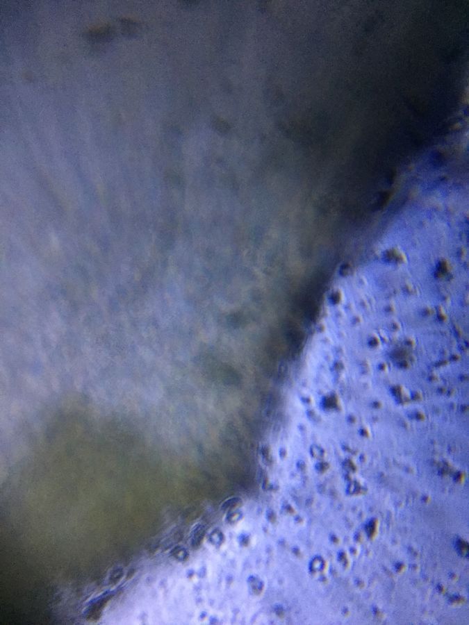
The following Foldscope images represent a persevering return to my samples from nearly seven weeks ago after an initially defeated attempt at operating these highly technical paper pieces of ingenuity (aka Foldscope). I won’t go into detail, but I think I may have figured out the strange smell that has been emanating from the fridge these past couple weeks. Amazingly, except for a bit of detrital slime on my sample of Mazzaella flaccida , with a little extra care, I was able to prepare slides and execute attempt number two, I think satisfactorily.
View in Media Gallery
Failed Porphyra perforata attempt 1.
View in Media Gallery
Failed Mastocarpus papillatus attempt 1.
View in Media Gallery
Failed Mazzaella flaccida attempt 1.
These images show that second frustrating Foldscope experience. As you can see, one can’t glean much from these images.
I know there is some saying about trying again if you fail… anyhow, the following series of images reveals, well, more information certainly, but, well, let’s have a look.
These images show that second frustrating Foldscope experience. As you can see, one can’t glean much from these images.
I know there is some saying about trying again if you fail… anyhow, the following series of images reveals, well, more information certainly, but, well, let’s have a look.
View in Media Gallery
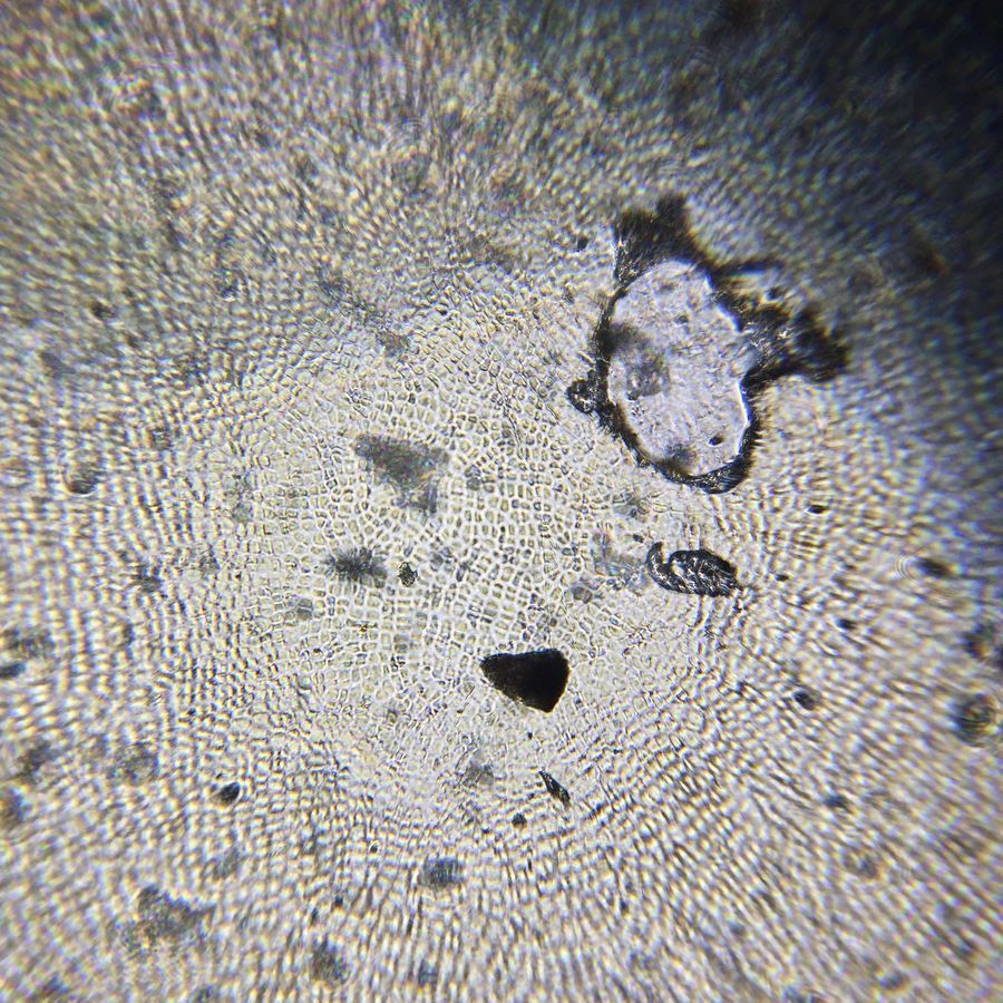
Porphyra perforata original slide preparation (dry).
View in Media Gallery
Porphyra perforata original slide preparation (dry).

View in Media Gallery
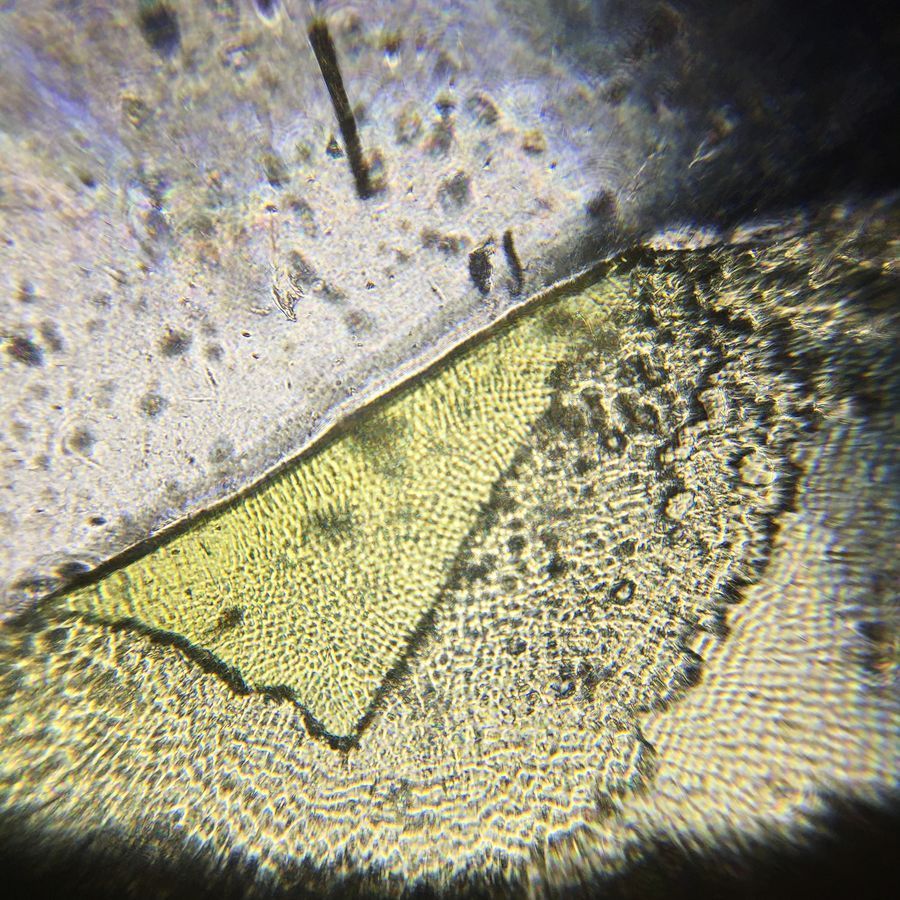
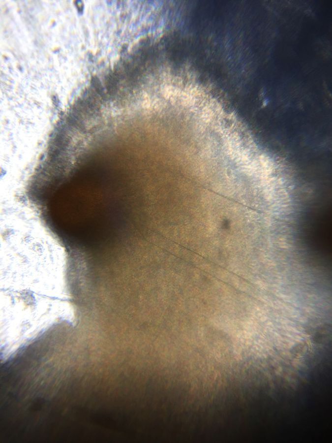
Mastocarpus papillatus cross section.
View in Media Gallery
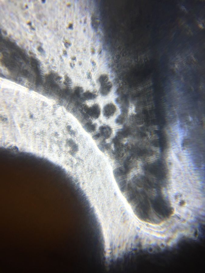
Mastocarpus papillatus, interesting.
View in Media Gallery
Mastocarpus papillatus cross section.
View in Media Gallery
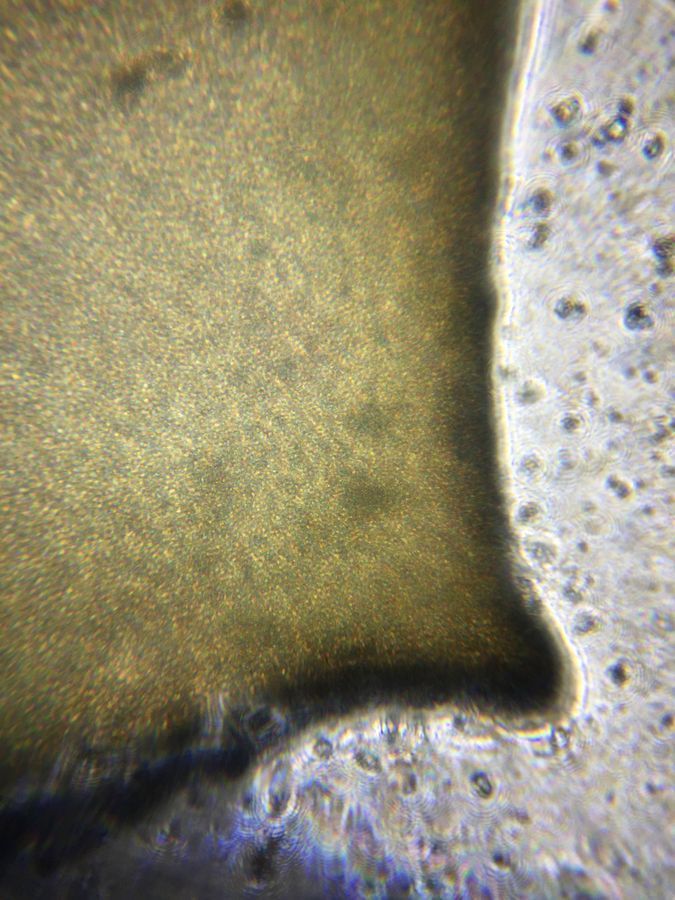
Mazzaella flaccida.

View in Media Gallery
Mazzaella flaccida, cross section?

View in Media Gallery
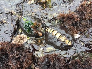
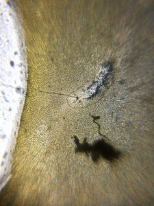
De-shelled little black snail found on Porphyra (already expired…in the fridge).
So, upon examining these images, I can’t say I’ve come much closer to uncovering any physical characteristics that would lead me to believe one of these species has any particular physical trait that could fend off desiccation or resist predation than another. However, we do see a few interesting differences. This Porphyra specimen appears to be composed of a single layer of cells, and it is quite thin to the touch. Mastocarpus blades are probably four to ten times thicker, and as we can see from the cross section, there is some interesting activity at the tip. Although I can’t put my finger on the exact description at present, I believe this is essentially the aggregation of supplies pursuant to cellular growth. Mazzaella displays a very uniform cellular pattern that appears many layers thick. The final slide shows some part of a small snail’s body (shell ~ 3mm long) that caught a ride in the Porphyra . Interestingly, aside from some linear membrane partitioning, the snail’s tissue appears to have a very similar texture to that of the seaweed it consumes?
Well, that’s going to have to do it for now. I haven’t given up on my investigation, and although I didn’t find what I was looking for, the journey has been well worth the effort. With a bit more time, I’m sure there is quite a bit more information in these images than I have touched upon. My interest and inspiration to trod on in my Foldscope journey has been renewed and I eagerly await my next adventure!
Sign in to commentNobody has commented yet... Share your thoughts with the author and start the discussion!
 0 Applause
 0 Comments