Macroalgal zonation, revisited
 May 14, 2017 • 9:41 PM UTC
 Unknown Location
 140x Magnification
 Plants
Shannon Myers
Learn about the author...
3posts
0comments
2locations
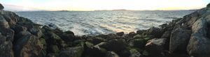
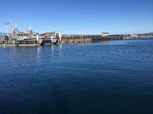
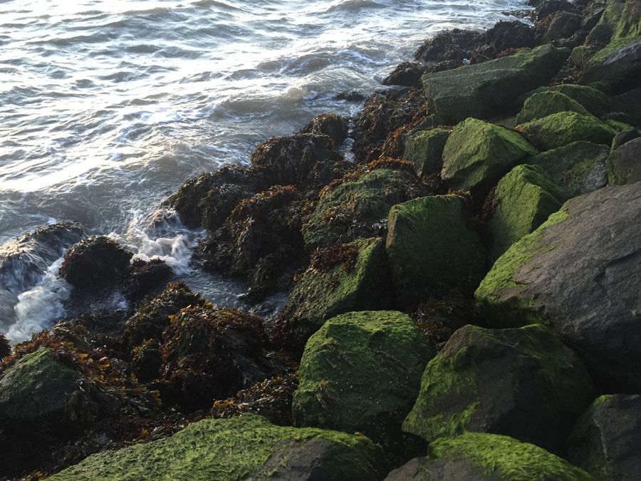
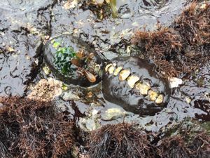
Hello, Shannon here with one more intertidal attempt at demystifying seaweed zonation patterns in a different environment, this time, the San Francisco Bay. Emeryville Marina Park is an elbow that juts out into the bay, and in all likelihood use to be part of the bay. Although this is an artificial rocky intertidal, and with significantly different physical and biological dynamics than the rugged open coastal location I previously visited, I spotted some similar species of macroalgae, and, a clear vertical zonation pattern. At this location, three zonally distinct species included (from the upper zone): Urospora penicilliformis (a bright green species with many long, hollow, filamentous strands draping over rock faces), Porphyra perforata (Nori), and then slightly interspersed Fucus gardneri and Mastocarpus papillatus . My objective at this site: obtain samples of these zonally characteristic species and try again with the help of microscopy to see any structural differences that might elucidate some facet of this pattern.
View in Media Gallery
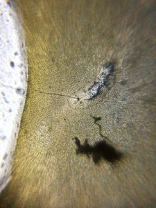
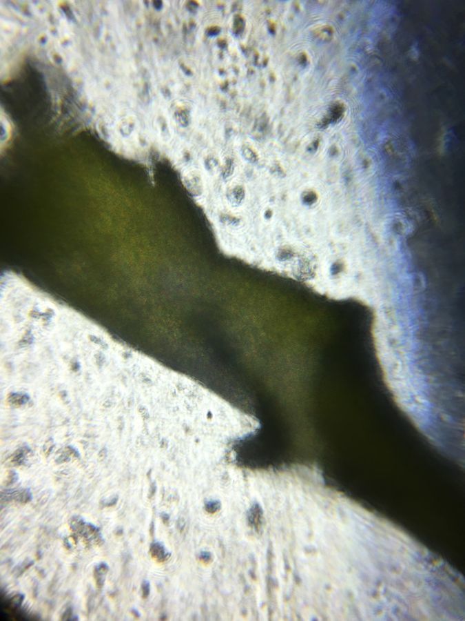
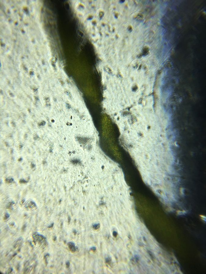
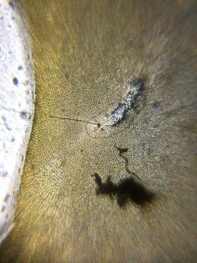
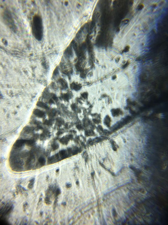
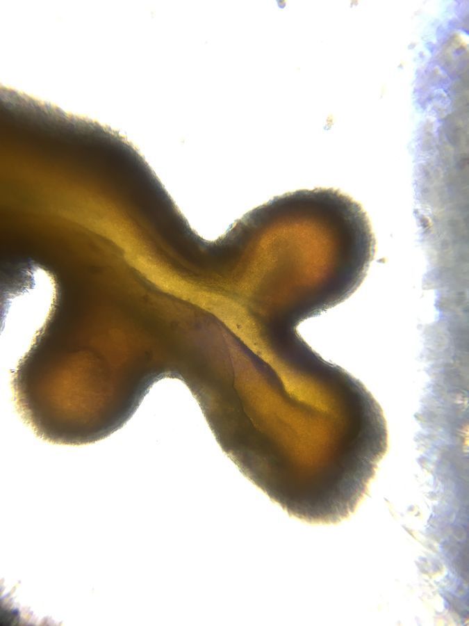
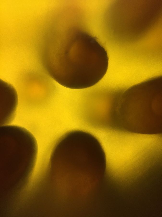
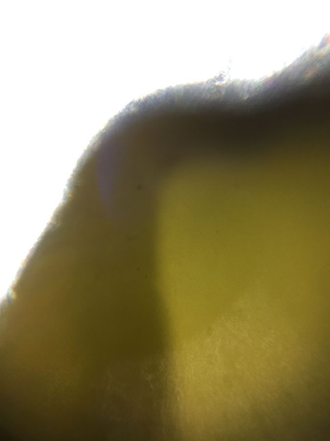
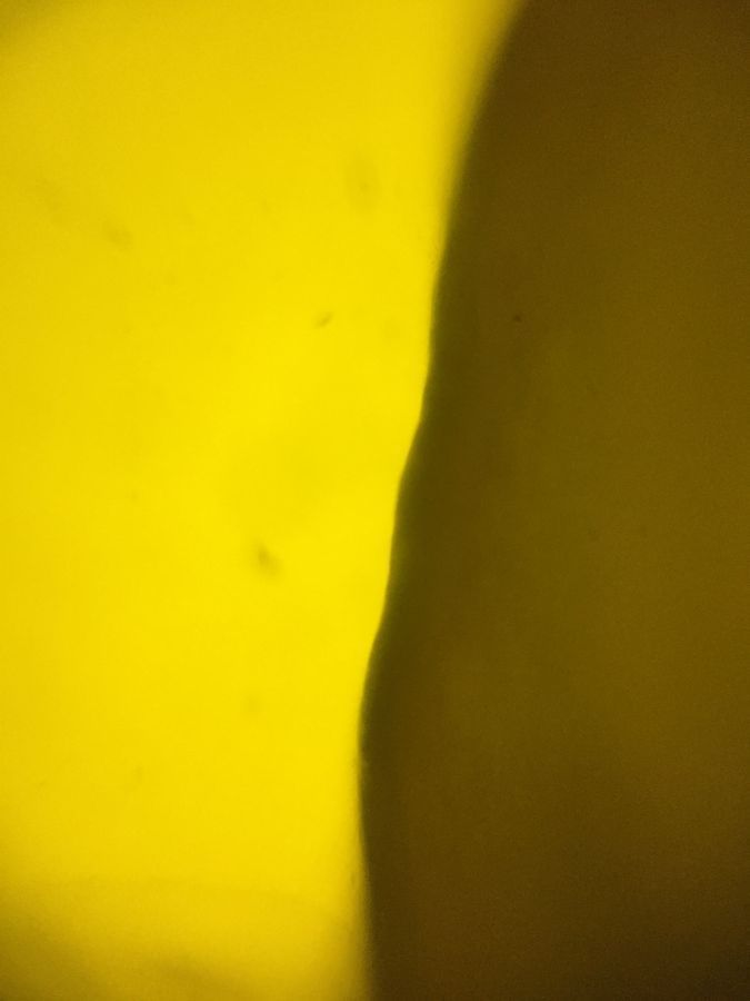
Here is what I came up with. Although I can’t quite make out distinct cells of Urospora , these hollow tubes appear to fold over and twist on themselves. In the left image, it appears these tubes have tiny openings that may be involved in the exchange of water or gases? Porphyra displays a cellular arrangement that is somewhat uniform and appears one dimensional. The cross section of Mastocarpus displays an aggregation of cellular materials near the distal extremities of the thallus. To be able to capture this cellular activity with a foldscope that costs less than a cup of coffee is truly remarkable! The next two images show differing forms of the papillae found on Mastocarpus papillatus . It is hard to see any cellular features, but what a fascinating morphology. I can’t say that the images of Fucus gardneri lend much insight into how this species makes a living in the lower intertidal, but its cells must be very small to form such a uniform-textured thallus.
View in Media Gallery
Urospora penicilliformis.
View in Media Gallery
Urospora penicilliformis.
View in Media Gallery
Porphyra perforata.
View in Media Gallery
Mastocarpus papillatus cross section.
View in Media Gallery
Mastocarpus papillatus.
View in Media Gallery
Mastocarpus papillatus.
View in Media Gallery
Fucus gardneri midrib slice.
View in Media Gallery
Fucus gardneri w/ partial midrib missing.
Although this was a lot of fun, I think in order to answer my original question as to whether there are any clear morphological characteristics that give one species a clear fitness advantage over another in the intertidal environment, I probably need to set down my Foldscope for a while and do some additional research. I am a big advocate for the transformative power that scientific literacy and interaction with nature can have on society; I strongly believe that if more people in this world took a little more time to get out and remember those early memories of wonder and awe at the incredible physical reality that surrounds us, maybe some of our choices would be a little different. Thank you Foldscope team for creating an incredible tool that I know will inspire countless minds to explore the small things in nature’s beautiful and infinite complexity. Until next time, have fun exploring everyone!
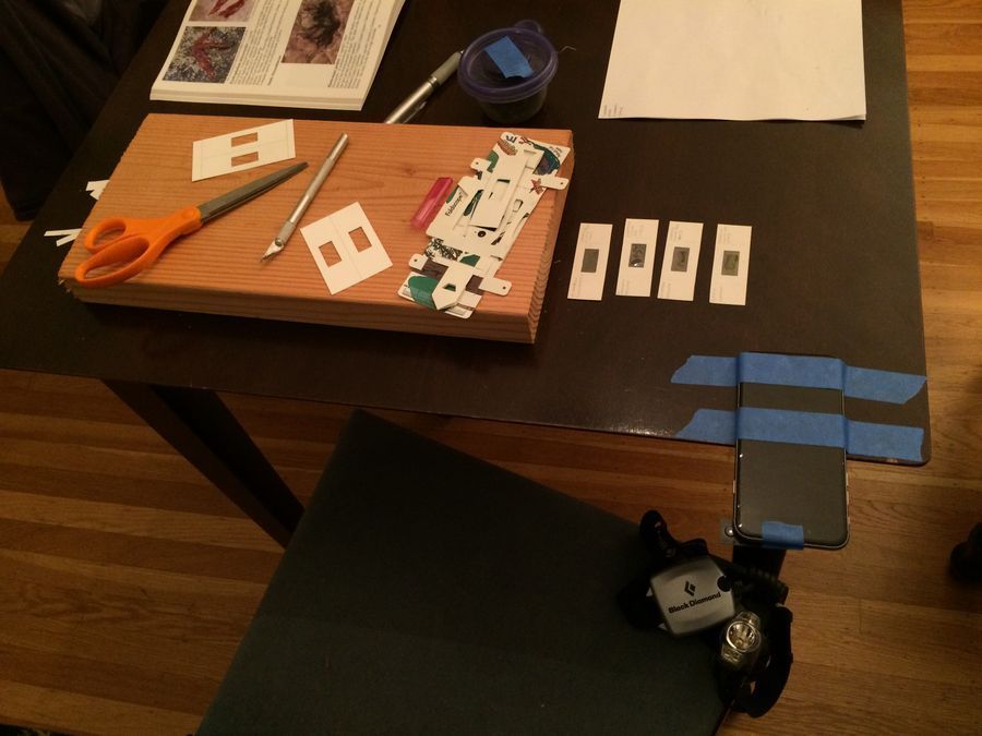
I nearly forgot to include a picture of my Foldscope workbench. Business cards make two slides perfectly, and with a block of wood, an x-acto knife, scissors, and a roll of packing tape, you will never run out of slides!
Although this was a lot of fun, I think in order to answer my original question as to whether there are any clear morphological characteristics that give one species a clear fitness advantage over another in the intertidal environment, I probably need to set down my Foldscope for a while and do some additional research. I am a big advocate for the transformative power that scientific literacy and interaction with nature can have on society; I strongly believe that if more people in this world took a little more time to get out and remember those early memories of wonder and awe at the incredible physical reality that surrounds us, maybe some of our choices would be a little different. Thank you Foldscope team for creating an incredible tool that I know will inspire countless minds to explore the small things in nature’s beautiful and infinite complexity. Until next time, have fun exploring everyone!
I nearly forgot to include a picture of my Foldscope workbench. Business cards make two slides perfectly, and with a block of wood, an x-acto knife, scissors, and a roll of packing tape, you will never run out of slides!
View in Media Gallery
Sign in to commentNobody has commented yet... Share your thoughts with the author and start the discussion!
 0 Applause
 0 Comments