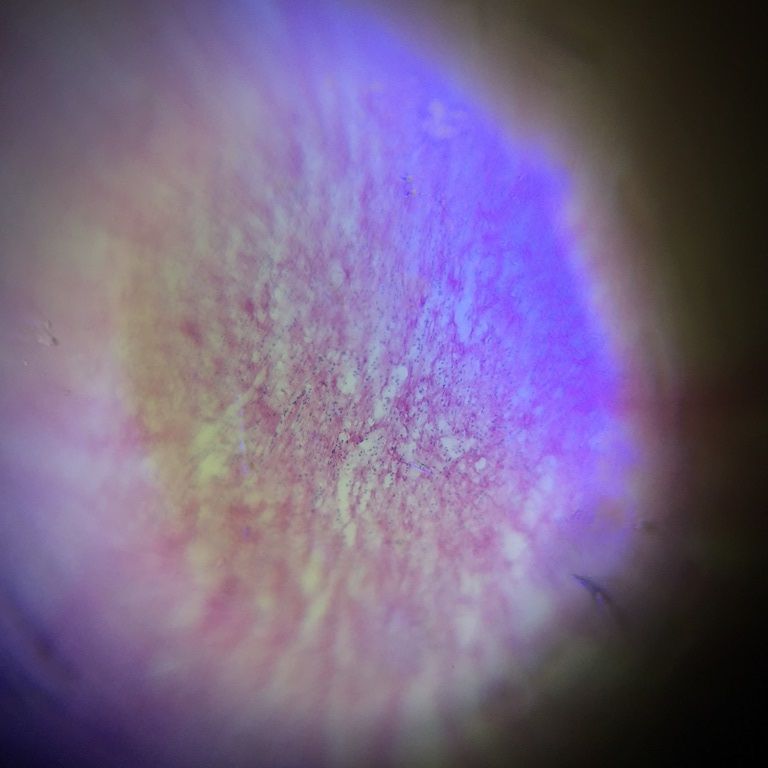
Chicken’s heart tissue
 Mar 06, 2015 • 12:44 AM UTC
 Unknown Location
 140x Magnification
 Microorganisms
Joelle Chang
I'm a lecturer in the Institute of Technical Education (ITE) in Singapore. Although I've been trained as a Chemist, but my interest in photography has taken me to embark on a Photomicrography Project with a Biology-trained colleague. Together with a few of our students, we have produced images of the microscopic world and have put up two annual exhibition in our institution. Here's the facebook album of our exhibits: https://www.facebook.com/media/set/?set=a.229241630578891.1073741829.100004791662049&type=1&l=ff618cd322
1posts
0comments
1locations
View in Media Gallery
Hello! This is my first post and my first time trying out the foldscope. Yesterday, my colleague prepared some slides of chicken’s heart tissue (sectioned using the cryostat). The image on the right is taken under Nikon Ci-L microscope (400x magnification), while the one on the left is taken using foldscope (low magnification). Both images were taken using iphone6. I noticed that there is always some bluish light in the foldscope picture, and sometimes, it appears in the middle. Any possibility of getting rid of it?
The dark purple spots are the nuclei, which are slightly visible even on low magnification. Unfortunately, I wasn’t able to get the focus using the high magnification lens.
The dark purple spots are the nuclei, which are slightly visible even on low magnification. Unfortunately, I wasn’t able to get the focus using the high magnification lens.


~ Joelle ~
Sign in to commentNobody has commented yet... Share your thoughts with the author and start the discussion!
More Posts from Joelle Chang
No more posts from this author.