Microbiology of Idli and Dosa
 Mar 12, 2015 • 2:14 PM UTC
 Unknown Location
 140x Magnification
 Microorganisms
Laks Iyer
Human observer of life. https://sukshmadarshin.wordpress.com
97posts
1255comments
5locations
View in Media Gallery
Prelude: I thought I was going to write a post on the microbiology of the Idli, but I worry this reads more like a cooking post. Apologies for the rambling, but I write for reproducibility and as a record for myself.
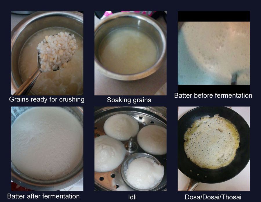
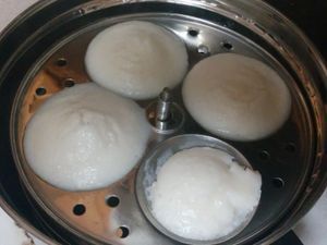
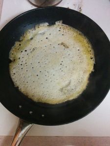
The Idli and Dosa (also Dosai/Thosai) are popular fermented foods that originated in the south of India and have spread world-wide. Both are derived from the same batter and every household in the Indian south prides itself about its idlis and closely guards its variations. The earliest mention of an Idli-like dish goes back some 1000 years and I suspect that the invention is likely to be older. The basic components of the modern idli are only two: urad daal (black mungo gram, Vigna Mungo) and rice (I use parboiled rice, Ponni type). The proportions of these matter for the quality of the product and I mix them in a 1:3 (1: Legume, 3: Rice), additionally people have their own additives such as fenugreek seeds and so on. My own recipe, passed down from hoary antiquity, is as follows: The grains are washed and soaked overnight (Figure 1 first row) and in the morning they are crushed in a grinder with water and salt added to taste (the differences between preparations lies in the fine art of knowing how fine/ coarse to grind the grains to get the right consistency). The batter is set to ferment for 12 hours or so at room temperature (above 25 C). Once it leavens it is poured into idli moulds and steamed to make soft and porous idlis or lightly fried in a pan to make the dosa (which looks like the western crepe) (Figure 1, second row). They have a slightly sour taste, a nice aroma (diacetyl?) and can be combined in a hundred ways with various chutneys and fillings.
The Idli and Dosa (also Dosai/Thosai) are popular fermented foods that originated in the south of India and have spread world-wide. Both are derived from the same batter and every household in the Indian south prides itself about its idlis and closely guards its variations. The earliest mention of an Idli-like dish goes back some 1000 years and I suspect that the invention is likely to be older. The basic components of the modern idli are only two: urad daal (black mungo gram, Vigna Mungo) and rice (I use parboiled rice, Ponni type). The proportions of these matter for the quality of the product and I mix them in a 1:3 (1: Legume, 3: Rice), additionally people have their own additives such as fenugreek seeds and so on. My own recipe, passed down from hoary antiquity, is as follows: The grains are washed and soaked overnight (Figure 1 first row) and in the morning they are crushed in a grinder with water and salt added to taste (the differences between preparations lies in the fine art of knowing how fine/ coarse to grind the grains to get the right consistency). The batter is set to ferment for 12 hours or so at room temperature (above 25 C). Once it leavens it is poured into idli moulds and steamed to make soft and porous idlis or lightly fried in a pan to make the dosa (which looks like the western crepe) (Figure 1, second row). They have a slightly sour taste, a nice aroma (diacetyl?) and can be combined in a hundred ways with various chutneys and fillings.
View in Media Gallery
Figure 1
The amazing thing is that this recipe has worked every time I made it with my clumsy hands without any external addition of microbes. This is because the normal flora of the Vigna mungo legume seed is self-sufficient and serves as the starter culture for fermentation and leavening. Studies have shown that the dominant microbes involved in this are lactic acid bacteria like Leuconostoc and Lactobacillus . However, being a mixed culture, the entire range of micro-organisms is not known and aspects such as the nature of microflora transitions should be studied given that a few million humans consume it almost every day. With modern sequencing technology, the idli batter is ripe for microbiome studies. I for one am sure that there are different types of starting bacteria and definitely non- Lactic acid bacteria as well. However, once the lactic acid bacteria start fermenting the batter and reduce the pH, the medium becomes inhospitable to the others. Moreover, we are only beginning to understand the role of inter-bacterial competition. Its a war zone out there and we use it to make our food.
My objectives were rather simple, I just wanted to foldscope the batter. Viewing bacteria at 140x and 400x especially those that dont move or have interesting structures can be rough, but I have not peered into the batter for long and so I took the plunge.
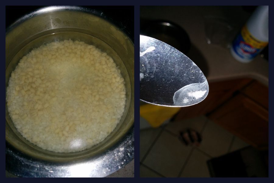
Below in Figure 2, on the left is shown the soaking grains before crushing. A nice biofilm of bacteria has already started to form after overnight soaking and I took a small spoonful. I hope you can see the bubbles suggesting the beginning of the fermentation process perhaps from the dissolved starch (The pH of the supernatent was about 6 at this stage).
The amazing thing is that this recipe has worked every time I made it with my clumsy hands without any external addition of microbes. This is because the normal flora of the Vigna mungo legume seed is self-sufficient and serves as the starter culture for fermentation and leavening. Studies have shown that the dominant microbes involved in this are lactic acid bacteria like Leuconostoc and Lactobacillus . However, being a mixed culture, the entire range of micro-organisms is not known and aspects such as the nature of microflora transitions should be studied given that a few million humans consume it almost every day. With modern sequencing technology, the idli batter is ripe for microbiome studies. I for one am sure that there are different types of starting bacteria and definitely non- Lactic acid bacteria as well. However, once the lactic acid bacteria start fermenting the batter and reduce the pH, the medium becomes inhospitable to the others. Moreover, we are only beginning to understand the role of inter-bacterial competition. Its a war zone out there and we use it to make our food.
My objectives were rather simple, I just wanted to foldscope the batter. Viewing bacteria at 140x and 400x especially those that dont move or have interesting structures can be rough, but I have not peered into the batter for long and so I took the plunge.
Below in Figure 2, on the left is shown the soaking grains before crushing. A nice biofilm of bacteria has already started to form after overnight soaking and I took a small spoonful. I hope you can see the bubbles suggesting the beginning of the fermentation process perhaps from the dissolved starch (The pH of the supernatent was about 6 at this stage).
View in Media Gallery
Figure 2
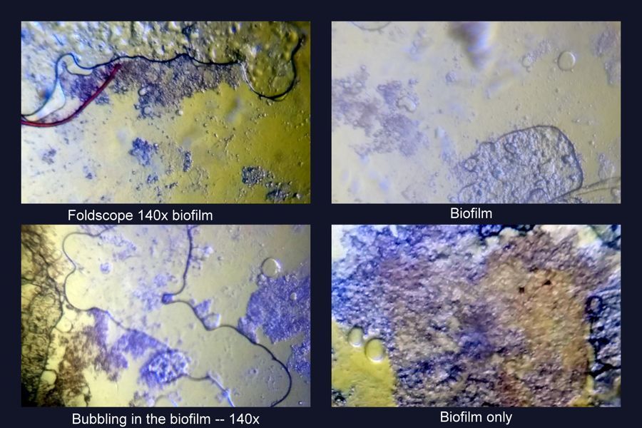
The biofilm is interesting under the foldscope. It was bubbling even as I was viewing it (Figure 3).
The biofilm is interesting under the foldscope. It was bubbling even as I was viewing it (Figure 3).
View in Media Gallery
Figure 3
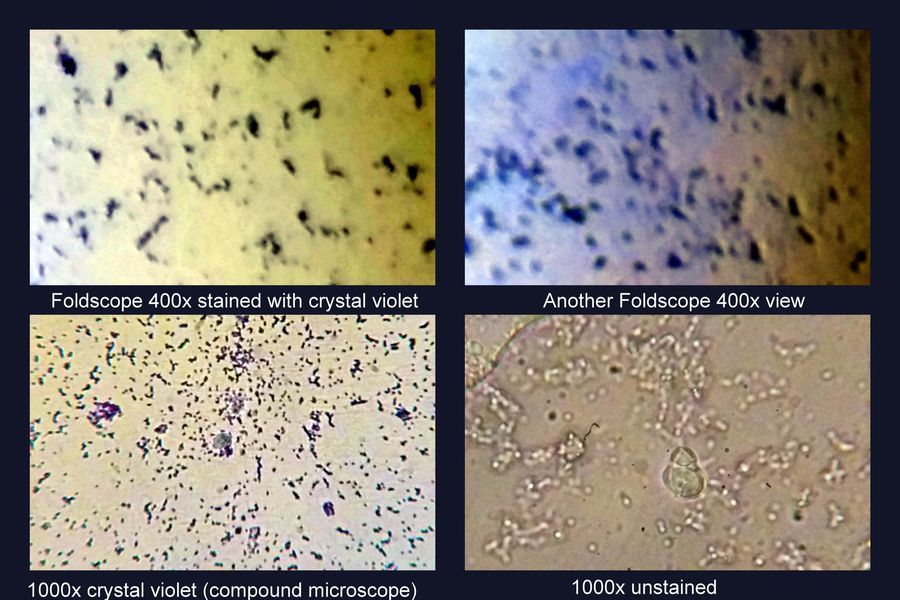
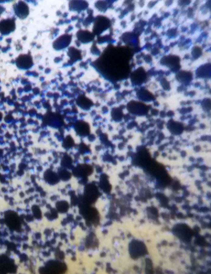
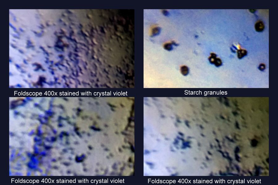
Given the transparency of bacteria and the small size of the Lactobacilli, I stained it to see their entire range of shapes and form. For this, I put a drop of the supernatent on a slide and let it air dry and then a drop of Crystal Violet for a minute and washed it off gently with water. The field is densely populated with various bacteria (Figure 4). Lactobacilli are likely to be the smaller ones, they are described as coccoid-ovoids and hence I include a couple of frames from my 1000x oil immersion compound microscope. The foldscsope high power however does show rods, cocci and some chains.
Given the transparency of bacteria and the small size of the Lactobacilli, I stained it to see their entire range of shapes and form. For this, I put a drop of the supernatent on a slide and let it air dry and then a drop of Crystal Violet for a minute and washed it off gently with water. The field is densely populated with various bacteria (Figure 4). Lactobacilli are likely to be the smaller ones, they are described as coccoid-ovoids and hence I include a couple of frames from my 1000x oil immersion compound microscope. The foldscsope high power however does show rods, cocci and some chains.
View in Media Gallery
Figure 4
I was greatly excited by watching motile bacteria in the supernatent under high power (400x). Bacteria tumble and run and it is delightful to see this (Figure 5, video). Note Lactobacillus and related bacteria are non-motile and hence the motile ones are likely to be other types of bacteria such as proteobacteria. Every time I see something under a foldscope, I keep telling myself, I wish I had one when I was 10. Also included in the below video at the end is a 1000x compound microscope shot that illustrates the size difference between the motile and non-motile bacteria.
I was greatly excited by watching motile bacteria in the supernatent under high power (400x). Bacteria tumble and run and it is delightful to see this (Figure 5, video). Note Lactobacillus and related bacteria are non-motile and hence the motile ones are likely to be other types of bacteria such as proteobacteria. Every time I see something under a foldscope, I keep telling myself, I wish I had one when I was 10. Also included in the below video at the end is a 1000x compound microscope shot that illustrates the size difference between the motile and non-motile bacteria.
Figure 5 (Video)
Post crushing the batter is flat and since there is likely to be a lot of starch due to the rice and the bean used in the preparation, I thought it might be interesting to see the particles of starch in the batter. Since starch easily stains with iodine (CVS brand), I added some into the batter, diluted it and put it on a slide (Figure 6). In this preparation, two sizes of particles can be seen, one is likely to be rice and the other the urad daal. Which one is which?
Post crushing the batter is flat and since there is likely to be a lot of starch due to the rice and the bean used in the preparation, I thought it might be interesting to see the particles of starch in the batter. Since starch easily stains with iodine (CVS brand), I added some into the batter, diluted it and put it on a slide (Figure 6). In this preparation, two sizes of particles can be seen, one is likely to be rice and the other the urad daal. Which one is which?
View in Media Gallery
Figure 6
Post-fermentation, there is a lot of bubbling of CO2 and the batter is leavened and thickened. The pH dropped to about 4. Here is a 7 minute shot shown at 16x the original speed (Figure 7, Video). The video shows the bubbles emerging underneath and getting trapped in the batter. You should pay attention all over the slide to see this. The thing is that the batter has to be thick enough to hold the CO2 bubbles. By this the density reduces by increasing volume and become fluffy upon cooking.
Post-fermentation, there is a lot of bubbling of CO2 and the batter is leavened and thickened. The pH dropped to about 4. Here is a 7 minute shot shown at 16x the original speed (Figure 7, Video). The video shows the bubbles emerging underneath and getting trapped in the batter. You should pay attention all over the slide to see this. The thing is that the batter has to be thick enough to hold the CO2 bubbles. By this the density reduces by increasing volume and become fluffy upon cooking.
Figure 7 (Video)
Since I thought it might be difficult to see the bacteria among starch granules,I diluted the batter many folds with 3 serial dilutions (I wasn’t too quantitative here) and stained it with crystal violet for foldscoping (Figure 8). The starch granules are actually easily disingtuished, they are refractile, transparent in the center with an outline. The motile bacteria were no longer see, instead the field was filled with very small bacteria.
Since I thought it might be difficult to see the bacteria among starch granules,I diluted the batter many folds with 3 serial dilutions (I wasn’t too quantitative here) and stained it with crystal violet for foldscoping (Figure 8). The starch granules are actually easily disingtuished, they are refractile, transparent in the center with an outline. The motile bacteria were no longer see, instead the field was filled with very small bacteria.
View in Media Gallery
Figure 8
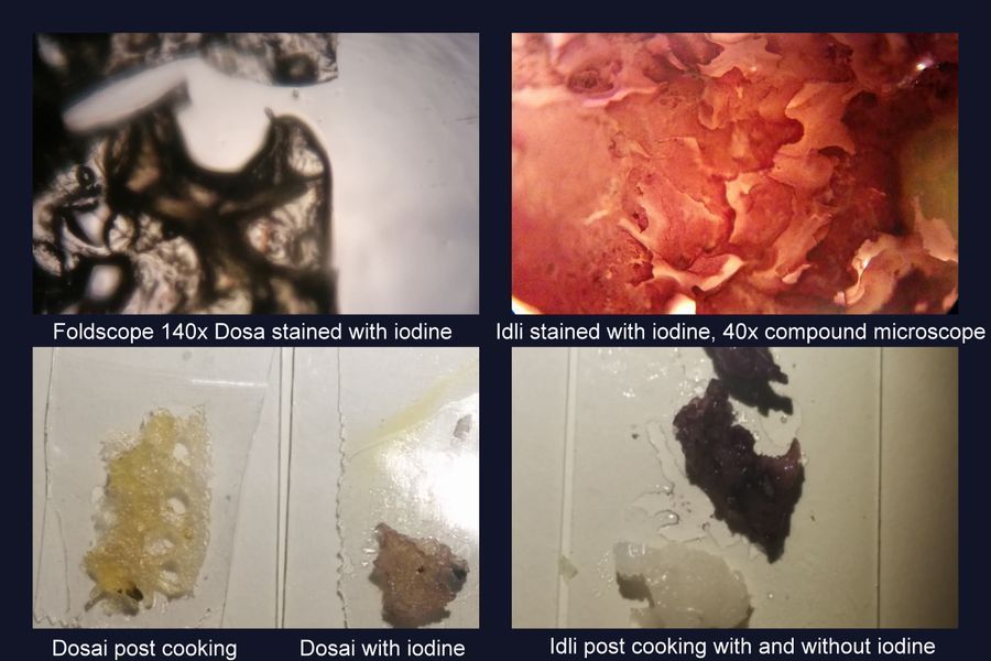
The steamed idli and pan-fried dosais are delightful dishes, but I wondered what they would look like under the microscope and so I took a small piece of each and stained it with dilute iodine (Figure 8). Below the dosai foldscoped well due to its porosity, not the idli for which I used a 40x compound microscope. The idli has some nice artistry. These and the regular picture shows that the idli or dosai is evently coated with starch. Upon heating the legume and rice proteins precipitate and also the starch into a CO2 filled matrix. Next time, I might try coomassie blue to stain the proteins.
The steamed idli and pan-fried dosais are delightful dishes, but I wondered what they would look like under the microscope and so I took a small piece of each and stained it with dilute iodine (Figure 8). Below the dosai foldscoped well due to its porosity, not the idli for which I used a 40x compound microscope. The idli has some nice artistry. These and the regular picture shows that the idli or dosai is evently coated with starch. Upon heating the legume and rice proteins precipitate and also the starch into a CO2 filled matrix. Next time, I might try coomassie blue to stain the proteins.
View in Media Gallery
Figure 9
Enough foldscoping for now, I cant wait to dive in when I see such pictures. Idlis are best eaten hot with a nice sambar and chutneys and also the thosai :). If you havent eaten it, you must try some. The post is dedicated to that pioneer/s who invented the idli and the dosai.
Comments, recommendations, criticisms always welcome.
Sign in to commentNobody has commented yet... Share your thoughts with the author and start the discussion!

 0 Applause
 0 Comments
_300x300.jpeg)


