First Experiments with Foldscope
 Dec 06, 2017 • 2:40 PM UTC
 Unknown Location
 140x Magnification
 Microorganisms
Nora Wolcott
I am an Undergraduate Lab Assistant at George Washington University, working for Dr. Arnaud Martin to conduct CRISPR experiments in butterflies.
1posts
3comments
1locations
View in Media Gallery
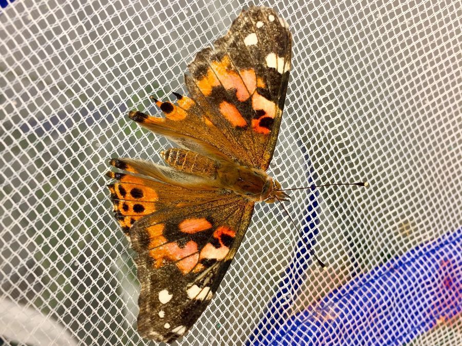
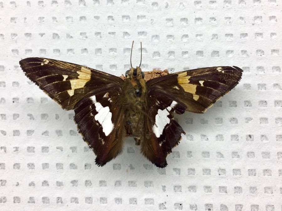
Today was my first attempt at using the foldscope to visualize scale structures, and overall I would consider it a success! Because most of my recent experiments have been focusing on Vanessa cardui , the painted lady butterfly, that was the species I began with. Putting together the foldscope I found the design quite clever and intuitive, simple enough that a resourceful child could put it together without too much difficulty. Vanessa , shown below, is a gorgeous, colorful species that has been fairly easy to raise in the lab, and I felt that the bright scales ranging from brown to pink to striking red, would be an excellent place to begin visualizing.
View in Media Gallery
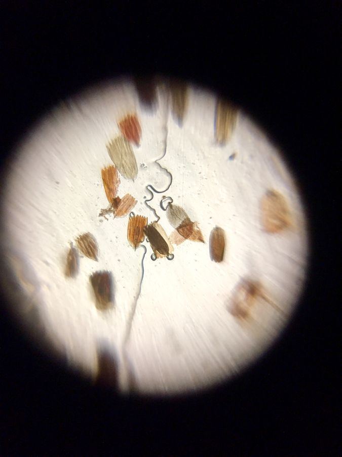
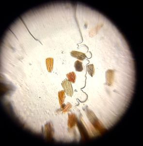
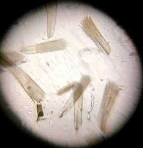
Using a small paintbrush I put a few scales onto a slide, and though my first attempt yielded too many scales (making it too opaque to visualize), my second went fairly well. As shown below, you can really see the variable color in the scales, and the complexity of the chitin that composes them. I was hoping to be able to visualize the many ridges in the scale cells, a goal which was partially accomplished, as you can see that the scales do not exhibit a flat surface. In the future I hope to collect slides with a smoother, more focused composition, and also reduce the amount of bubbles captured in the slide.

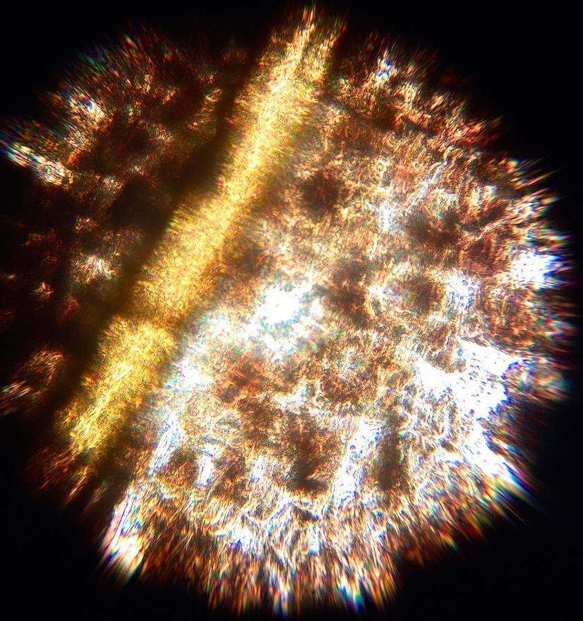
After this experiment went well, I then endeavored to visualize some thicker structures. Some, like legs and antennae, were irreparably opaque. Possibly in the future I can put together a method for stripping thicker structures down to a more transparent membrane, while retaining the composition of the structure (i.e. the hooks in the leg, the pigment of the antennae, etc.). One more successful thing I attempted was stripping a segment of a wing from a dead butterfly, who’s tissue and scales had been reduced to a very thin, delicate layer. Under normal lighting it was still too opaque, but backlit but a microscope light it resulted in a quite striking, kaleidoscopic image, seen below. While you can’t quite make out the scale structures, the vein running through the wing makes the image quite compelling.
View in Media Gallery
The second species I worked with today was Epargyreus clarus , the silver spotted skipper, which has the unique phenotypic attribute of iridescent, reflective scales on the hindwing (see below). Taken from a pinned specimen, I was interested in seeing the structure and transparency of these scales as opposed to the more heavily pigmented Vanessa.
View in Media Gallery
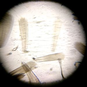
Once under the foldscope, the first thing that really struck me was the length of the scales. You can clearly see that, as compared to the scales of Vanessa, they are roughly twice the length. I think that moving forward it would be interesting to see if the length contributes to the iridescent nature of the scales, and if it is echoed in the brown pigmented sections of the wing. This also raises evolutionary questions too broad to answer in the timespan I have, concerning the interrelatedness of Vanessa and Epargyreus. The lack of pigment also made the scales slightly difficult to get in focus, something that I have been having a bit of trouble with all around. I think that as I continue to work with the foldscope, the Z scale should come more naturally to me.
Overall, I would consider today quite a success! The concept of the scope as a way to get microscopes into the hands of children across the world is a really amazing one, and a goal I think is absolutely achievable with such innovative technology. I know one thing, if I had had this as a kid I wouldn’t have been able to keep my hands off it. This is definitely a step up from the magnifying glass I carried with me through grade school. In the next couple weeks I hope to generate some mutant CRISPR embryos, which would be quite a step up, so stay tuned for some interesting developments!
Sign in to commentNobody has commented yet... Share your thoughts with the author and start the discussion!
More Posts from Nora Wolcott
No more posts from this author.