Slices of plants
 Dec 11, 2017 • 3:09 AM UTC
 Unknown Location
 140x Magnification
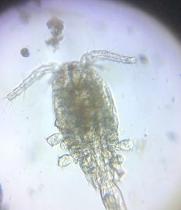
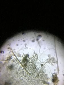
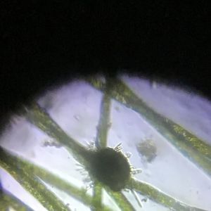
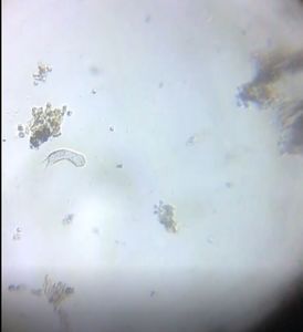
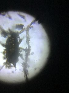
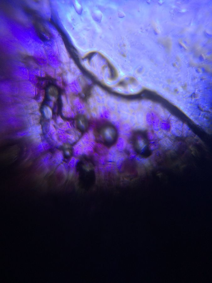
 Microorganisms
Jayashree Ramadas
We are a group of students, volunteers and staff working with TIFR Hyderabad's Science Education and Outreach program: http://www.tifrh.res.in/~outreach/
39posts
26comments
2locations

View in Media Gallery
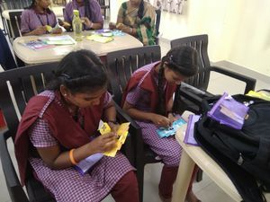
After our first ‘Atoms to Amoeba’ workshop four of our neighbouring TSWREIS (social welfare residential) schools took back two foldscopes each. Too few, yet so far several hundred students, most from remote rural parts of Telangana, have got their own “wow” moments on these foldscopes. It happened because our teacher participants went beyond conducting foldscope sessions for their own students. They pooled their eight foldscopes to set up a stall at the State-wide “Spectrum 2017” at Narsingi, which was attended by almost 1000 TSWREIS students from schools around Telangana. Foldscopes contributed by kind-hearted friends and colleagues have seeded this activity. If we could now get enough foldscopes to schools, we may see some real science projects at the 2018 festival.
View in Media Gallery
Foldscope stall at Spectrum 2017
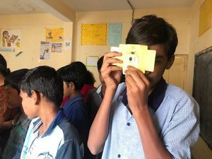
View in Media Gallery
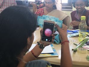
Trying foldscopes at Spectrum 2017. The girls on the right are demonstrating. Despite the challenges underlined in our last post we do hope to get foldscopes introduced into regular classroom teaching, lab sessions and students’ independent explorations. TSWREIS has 238 schools and they recently started 30 residential degree colleges for women, including special colleges for physical and life sciences. Young women arrive here from interior Telangana eager to learn, even while their labs and other infrastructure are still in the making. Driven by sheer motivation, they do rather well . Foldscopes would be great for them.

View in Media Gallery
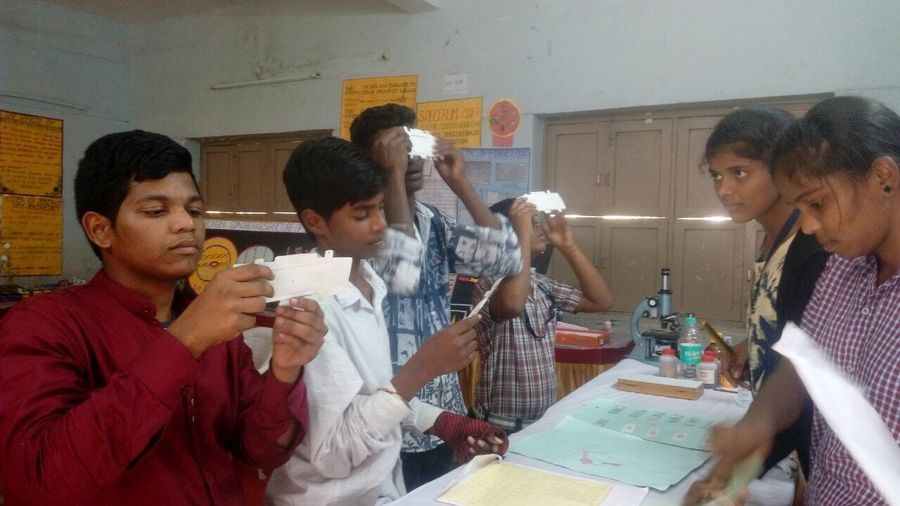
Students at life sciences college of TSWRDCW, Shamirpet To get foldscopes to students of government schools one must enter through the “practical” part of their curriculum. Searching for such singular openings, we found ourselves exploring plant tissues. TSWREIS students of Grade 11 at Gowlidoddi got really excited gathering, slicing and viewing sections of various stems, roots and leaves in their school grounds. Some of these students are here after doing Grade 10 in remote rural areas of Telangana. They got to see and handle their first dissection and compound microscopes, along with the foldscopes.
View in Media Gallery
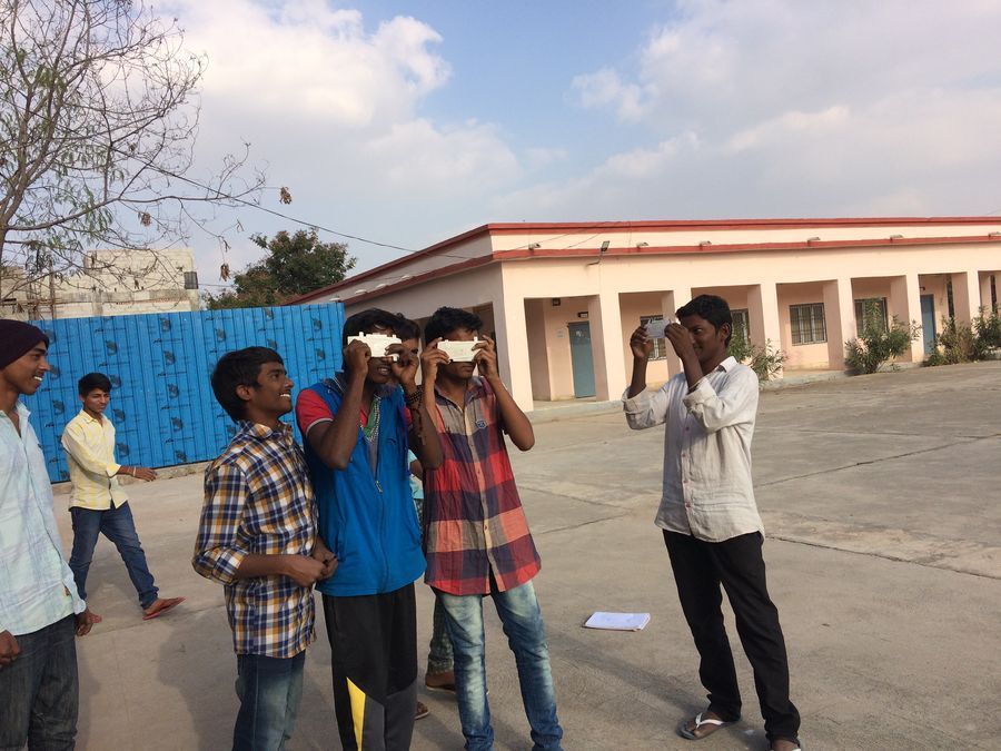
Plant tissues session at Gowlidoddi Boy’s school Slicing thin sections out of roots, stems and leaves was a challenge. But the razor blade feel of one-cell thickness does reward one with a rare, embodied sense of tens of microns.

View in Media Gallery
Relating textbook with experience Our first attempt with stems and roots of monocot grasses growing wild was disappointing. They seemed either too thin or too hard to get really good sections. (Later we had better luck with young bamboo shoots and roots.) For now we settled for commercially available preserved and enzyme-softened roots of Zea mays (corn) — which had lain in the lab, bottled and unopened for a year, perhaps two. The samples looked beautiful stained with saffranin, but once out on the slide the outer layer of cells disintegrated slowly. Here is our picture, air bubbles and all, but nicely showing large metaxylem vessels, part of a ring of smaller protoxylem vessels around them, phloem in between and pith at the centre. Here is a link to a much better picture with explanation.

View in Media Gallery
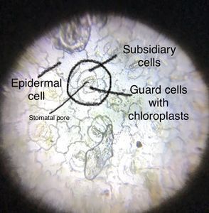
TS of Zea mays root This is the lower epidermis of a leaf of Rhoeo discolor (Tradescantia spathacea), in natural colour, showing black-lined stomata.
View in Media Gallery
Lower epidermis of Rhoeo discolor These are three images of TS of a leaf of a dicot plant that, the botany teachers among us guess, may be from the Annonaceae family. It is stained with saffranin.

View in Media Gallery
TS of dicot leaf midrib

View in Media Gallery
TS of dicot leaf midrib leading to lamina

View in Media Gallery
TS of dicot leaf lamina The first image shows the midrib part with the xylem. The second leads into the lamina of the leaf and the third is entirely the laminar part with what we guess is palisade parenchyma on the top and spongy parenchyma below. Please correct us if you know better! We are using the old model of the foldscope with a low magnification lens.
It is December already and our 11th and 12th graders are gearing up for their all-consuming ‘Board Exams’. So next we’ll use the foldscope with younger or older students. We’ll keep you posted.
— Jayashree Ramadas
It is December already and our 11th and 12th graders are gearing up for their all-consuming ‘Board Exams’. So next we’ll use the foldscope with younger or older students. We’ll keep you posted.
— Jayashree Ramadas
Sign in to commentNobody has commented yet... Share your thoughts with the author and start the discussion!

 0 Applause
 0 Comments