Exploring a Slime Mold-1: Detection, isolation and culturing
 Jan 19, 2018 • 9:36 AM UTC
 Unknown Location
 140x Magnification
 Microorganisms
Laks Iyer
Human observer of life. https://sukshmadarshin.wordpress.com
97posts
1255comments
5locations
View in Media Gallery
Slime molds have fascinated me from childhood. I read about them in a library book and as an undergrad tried to culture them with little success (all I got were tracks of gnats). The disappointment lasted many decades. A couple of years ago I got a sample of a slime mold from a friends’ porch, but they had bleach treated it (a name such as dog’s vomit doesn’t help), so it was essentially dead. Over the Christmas break of 2017, I read a fascinating book on slime molds (also called myxomycetes) by Steve Stephenson, Myxomycetes a Handbook of Slime Molds . and as luck would have it a slime mold found me, right in my own little garden. This time I was prepared and in the next 2-3 posts I shall outline my experiments with this mold and convince you that slime molds make great pets and are very amenable to microscopic study. But first what are slime molds?
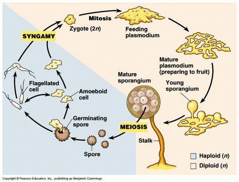
Slime molds or myxomycetes and their life cycles. Slime molds or myxomycetes are a fascinating branch of life. They are not a fungus (although they have the word mold in them), but like fungi they can fuse with one another (anastomose) or form fruiting bodies. They are closely related to amoebae such as Chaos , Amoeba proteus, and Entamoeba and most slime molds form a group with them called the amoebozoans. In the tree of life, amoebozoans form a branch outside animals and fungi. The three put together are often called unikonts . Now slime mold-like organisms are found in another branch of the tree of life ( acrasid slime molds ), but that is for another day. What makes the slime molds fascinating is that they have a unicellular phase and a multicellular phase. Take the example of a typical plasmodial slime mold like Physarum polycephalum (Figure 1).
Slime molds or myxomycetes and their life cycles. Slime molds or myxomycetes are a fascinating branch of life. They are not a fungus (although they have the word mold in them), but like fungi they can fuse with one another (anastomose) or form fruiting bodies. They are closely related to amoebae such as Chaos , Amoeba proteus, and Entamoeba and most slime molds form a group with them called the amoebozoans. In the tree of life, amoebozoans form a branch outside animals and fungi. The three put together are often called unikonts . Now slime mold-like organisms are found in another branch of the tree of life ( acrasid slime molds ), but that is for another day. What makes the slime molds fascinating is that they have a unicellular phase and a multicellular phase. Take the example of a typical plasmodial slime mold like Physarum polycephalum (Figure 1).
View in Media Gallery
Figure 1. Plasmodial slime mold life cycle. (Image from https://snowbio.wikispaces.com/Slime+Mold+%28protist%29) Starting from 6 ‘clock and going clockwise, haploid spores from the mold develop to give amoeboid or flagellated cells, which fuse to form a diploid organism, which further undergoes mitosis to form a plasmodium. Now many such plasmodia can fuse together to form a giant plasmodium, which contains a single cell membrane harboring 1000s of nuclei undergoing synchronous divisions. This feeding plasmodium forages and feeds and when resources dwindle, the nuclei are packaged as spores into the sporangium and thus a full life cycle is complete.

View in Media Gallery
Figure 2: Dictyostelium discoideum life cycle, picture from http://www.metamicrobe.com/dicty/ In contrast, in cellular slime molds like Dictyostelium (Figure 2, popularly called the social amoeba), the cells do not fuse, they just aggregate to form a slug-like form called a grex, which gives rise to fruiting bodies. What fascinating life cycles! To give you an analogy, imagine that each of your cells form separately from the other, forage and feed and then fuse at an appropriate time to form you and then you disintegrate to your individual cells and start afresh. This peculiar life cycle inspires many interesting theoretical problems in biology, like what determines if a cell or a nucleus become a spore (propagation into the next generation)? Are there cheater nuclei/cells that always become spores ? Could fusions occur between unrelated slime molds and how is that prevented? How do they deal with infections by viruses and bacteria? What molecules facilitate communication between the cells before fusion and after fusion, How is communication possible within the giant plasmodium… endless and fascinating questions, but for us with foldscopes our first goal is to see them.
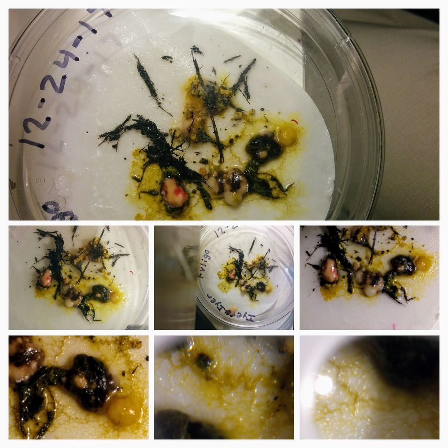
A slime mold in my garden mulch And so while I was reading the book, I decided to mulch my little garden for winter and lo and behold there was this yellow growth in the mulch (Figure 3A). Examination under the hand lens suggested that they were not fungi (no mycelia) but were likely to be a slime mold (Figure 3C/D). Figure 3C and D show magnifications of 3x and 10x taken with the lens-light provided with the new foldscope kit. Pot shaped sporangia, that were very crunchy to crush were visible. I crushed a bit of them to release spores (Figure 3B).
A slime mold in my garden mulch And so while I was reading the book, I decided to mulch my little garden for winter and lo and behold there was this yellow growth in the mulch (Figure 3A). Examination under the hand lens suggested that they were not fungi (no mycelia) but were likely to be a slime mold (Figure 3C/D). Figure 3C and D show magnifications of 3x and 10x taken with the lens-light provided with the new foldscope kit. Pot shaped sporangia, that were very crunchy to crush were visible. I crushed a bit of them to release spores (Figure 3B).

View in Media Gallery
Figure 3, A. Slime mold on mulch, B. Spores under a microscope, C.D: slime mold under the hand-held lens Here is a video of the spores with angular and full illumination.
I tried germinating the spores under a foldscope, with little success and so I tried culturing them. Luckily in the myxomycete book, I came across what is called the moist chamber technique. Here you put a couple of moist filter papers in a petridish (paper towels are also ok) and put barks or leaves on the wet paper for a few days until you start seeing telltale patterns of slime mold growth. I thought, I’d use the same technique but put some mulch with fruiting bodies (sporangia) in it and also oats as I read that certain plasmodial slime molds love oats(!) (Figure 4 A). And then I waited for something to happen, checking it regularly, either under a low power microscope (40x, Figure 4D-F), or under a foldscope (Figure 4B, note reflecting light hack). After about 10 days, and to my utter horror, I noticed that there was a fungus growing on it (Figure 4 D-F), although the fruiting body form had slowly dwindled. I thought that I was done in again.

View in Media Gallery
Figure 4 A. Set up for moist chamber, B. Slime mold as it looked after about a week under a foldscope. C-E Fungus growing on my slime mold After about 17 days as I was ready to throw my plate away in frustration, when I saw this.
View in Media Gallery
Figure 5. Plasmodium form of slime mold The fruiting bodies disappeared and there were these yellow streams and blobs all over the plate, indicative of an active plasmodium. The fungus also seemed to have grown a bit (colored), but the plasmodium was aggressively growing. Notice that although the pictures were taken on the same day, no two views are exactly alike. So I did a time lapse for about 2 hours and 40 min. taking a picture of the plate every 90 seconds and you can see the plasmodium explore the plate… (to be continued)
Sign in to commentNobody has commented yet... Share your thoughts with the author and start the discussion!

 0 Applause
 0 Comments
_300x300.jpeg)


