Two Types of Salt Crystals
 Feb 04, 2018 • 11:13 PM UTC
 Unknown Location
 140x Magnification
 Unknown
Mingqian Tan
Learn about the author...
2posts
0comments
1locations
View in Media Gallery
Students who are first learning how to use a microscope as part of an introductory science class often view salt or sugar crystals with the instrument–it’s a great and harmless way to introduce them to the microscopic world. Unfortunately, despite having worked with microscopes before I had yet to view salt crystals under one with my own eyes.
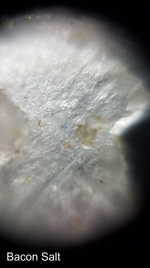
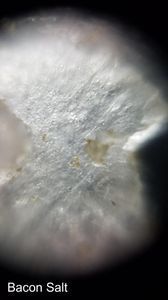
My roommate and I (both Bio 60 students this quarter) visited the Spice Library at the Arrillaga Family Dining Center to obtain samples of various spices. This post is about the two salt samples that we collected and viewed under our Foldscopes: smoked salt and bacon salt. The pictures are labeled with the corresponding type of salt.
My roommate and I (both Bio 60 students this quarter) visited the Spice Library at the Arrillaga Family Dining Center to obtain samples of various spices. This post is about the two salt samples that we collected and viewed under our Foldscopes: smoked salt and bacon salt. The pictures are labeled with the corresponding type of salt.
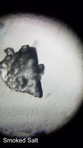
Both of the salts were viewed using dry mounts, as salts are ionic compounds that dissolve in water. Using tweezers, a small amount of salt crystals were sprinkled onto a clean microscope slide; a cover glass slip was then placed on top and sealed with tape. The slide was inserted very carefully and viewed with the Foldscope oriented horizontally, as the width of the salt crystals prevented a complete seal from the cover glass and so could fall out of the microscope slide if not handled carefully.
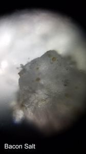
From the Foldscope exploration, both salts have the same general structure and appear as flattened, pointy flakes (the flatness is probably due to the pressure of the cover glass forcing the crystals to lie on their side).
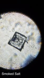
However, there was a particularly well-organized cubelike crystal in the smoked salt, which is included in this post.
The similarity of the crystals between the two salt mixes suggests that the unique flavors of each salt are due to other compounds mixed into the seasoned salt instead of differences inherent to the salt itself. I suspect the brown tinges in the bacon salt may be fat residues due to bacon bits (the bacon salt was simply a mix of normal salt and pieces of real bacon).
(Post is part of the #Bio60_2018 course.)
From the Foldscope exploration, both salts have the same general structure and appear as flattened, pointy flakes (the flatness is probably due to the pressure of the cover glass forcing the crystals to lie on their side).
However, there was a particularly well-organized cubelike crystal in the smoked salt, which is included in this post.
The similarity of the crystals between the two salt mixes suggests that the unique flavors of each salt are due to other compounds mixed into the seasoned salt instead of differences inherent to the salt itself. I suspect the brown tinges in the bacon salt may be fat residues due to bacon bits (the bacon salt was simply a mix of normal salt and pieces of real bacon).
(Post is part of the #Bio60_2018 course.)
Sign in to commentNobody has commented yet... Share your thoughts with the author and start the discussion!
 0 Applause
 0 Comments