A Blade of Grass (BIO60_2018)
 Feb 12, 2018 • 10:16 PM UTC
 Unknown Location
 140x Magnification
 Unknown
Annie Chang
Learn about the author...
2posts
0comments
1locations
View in Media Gallery
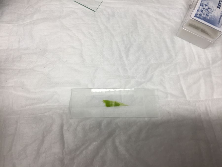
Nature abounds at Stanford. Outside the Gilbert Biology Building, for instance, is a bush of grass strands. I was curious what the inside of grass looks like. After plucking a strand out of a bush, we sliced it thinly with a razor so that we could examine its insides.
View in Media Gallery
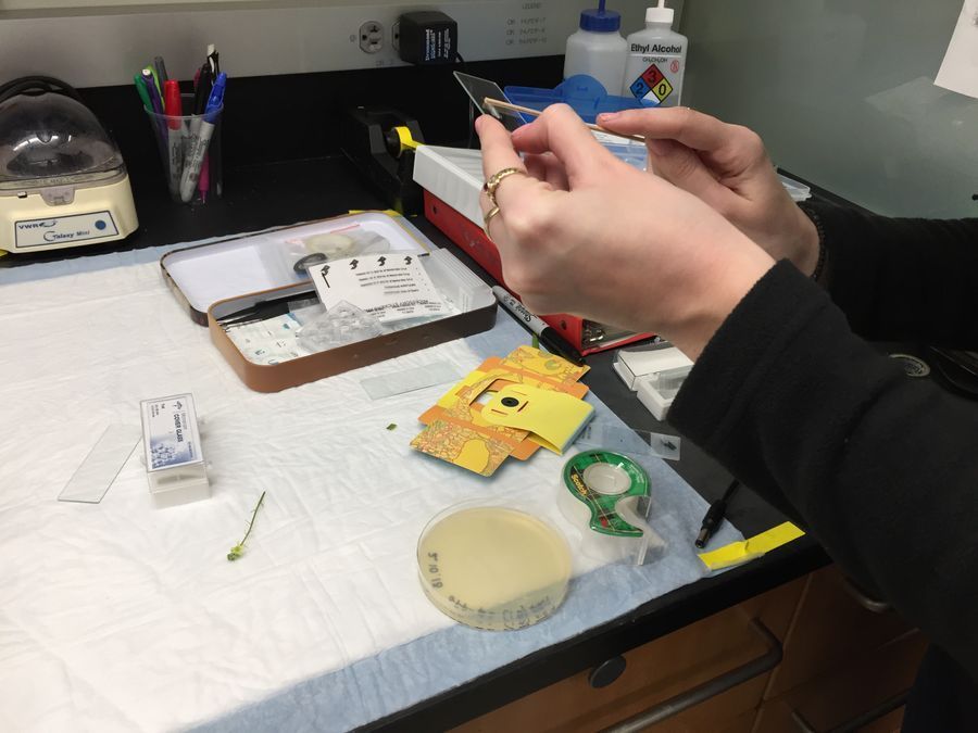
The process of preparing the grass slide involves slicing the grass, peeling it from the parent strand, and taping it to the slide. We also tried to look at E.coli bacteria (see below image of spreading Agar onto a cover glass), but the bacteria were too small to be seen clearly, and a small flower bud, which was too thick and not transparent enough.
View in Media Gallery
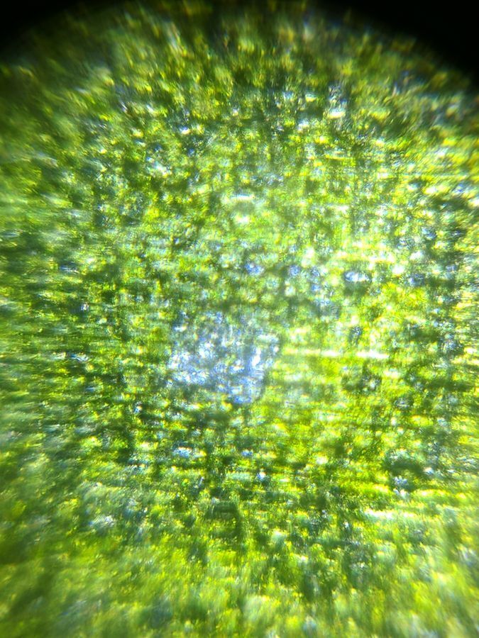
Lo and behold! After a few attempts, we were able to peer inside the grass. Here we can see the individual chloroplasts of the grass strand. These are visible because plant cells are larger than bacterial cells. I was surprised at the variety of green colors under the Foldscope, ranging from dark green to light green to yellow. The chloroplasts looked textured and rough, lining up closely next to each other in rectangular cell shapes to maximize the space available.
View in Media Gallery
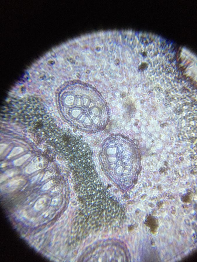
The image below is a view of an apical bud that was visible under the Foldscope. Here we can see cellular structures, perhaps vesicles, that are inside the bud. It appears that the bud is stained in different colors so that we can see the structures clearly. I wonder what the long, irregularly shaped green structure is.
View in Media Gallery
In the future, I’d like to look at yeast bacteria under the Foldscope.
Special shout out to Eirini for her help!
Bio60_2018
Special shout out to Eirini for her help!
Bio60_2018
Sign in to commentNobody has commented yet... Share your thoughts with the author and start the discussion!

 0 Applause
 0 Comments