Warm-Colored Flowers!
 Feb 24, 2018 • 12:46 PM UTC
 Unknown Location
 140x Magnification
 Unknown
Sandra Kong
Learn about the author...
2posts
0comments
1locations

View in Media Gallery
This week, as I was biking around campus, I noticed that flowers of many colors were starting to bloom! I was intrigued as to whether certain pigments would appear better in the foldscope and which types of petals had the most complexity. Unfortunately I had some difficulty viewing some of the other flowers I had collected around campus, probably due to their thickness, but I did manage to get pictures of some interesting warm-colored flowers.
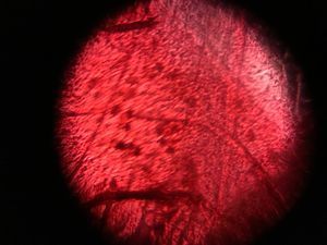
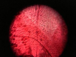
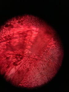
These are images of a red flower I collected outside my dorm. I wet mounted this sample, which helped a lot with viewing the petal. I believe the white shape at the top of the first two pictures is actually the light inside the classroom.

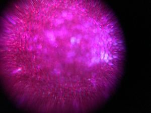

These are images of a magenta and white flower that was available in class. At one point I managed to position the slide so that I was able to see both white and magenta pigments. The pictures are a little blurry, but you can still see the cells that make up the petal pretty well! I found it interesting that the components in these pictures look quite different from the red flower’s pictures; however, I did mount the red flower a week before taking pictures, which may have affected the cells.
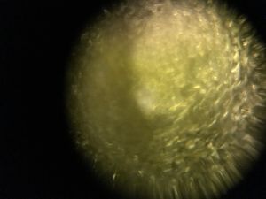
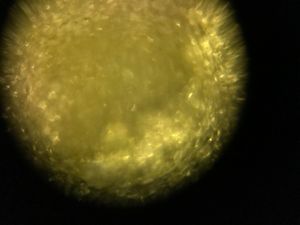
For this yellow flower, the petal alone was too thick to view, even with a wet mount, so I tried to scrape samples off the petal and wet mounted them. It seems like all you can really see is the yellow pigment over the bubbles of tape, but it’s still pretty cool!
Sign in to commentNobody has commented yet... Share your thoughts with the author and start the discussion!

 0 Applause
 0 Comments