Fungus on onion
 Mar 01, 2018 • 3:48 AM UTC
 Unknown Location
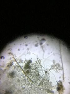
 140x Magnification
 Microorganisms
Jayashree Ramadas
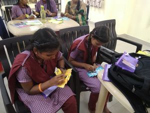
We are a group of students, volunteers and staff working with TIFR Hyderabad's Science Education and Outreach program: http://www.tifrh.res.in/~outreach/
39posts
26comments
2locations
View in Media Gallery
Onions in India often grow a black powdery fungus. Here’s a folscope-look at this fungus and the cells that it grows on.
View in Media Gallery
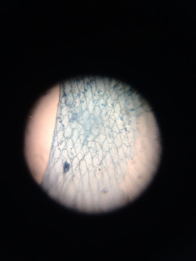
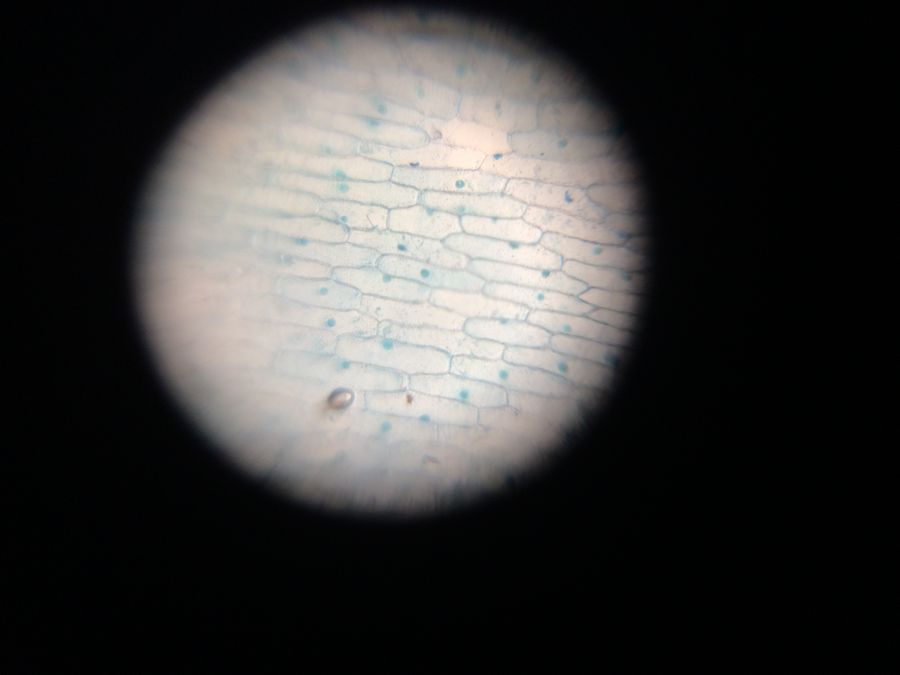
White onion with fungus First try – we discarded the dry papery outer peels and looked at the inner wet peel of the onion which was spotted with fungus. We stained it with methylene blue.
View in Media Gallery
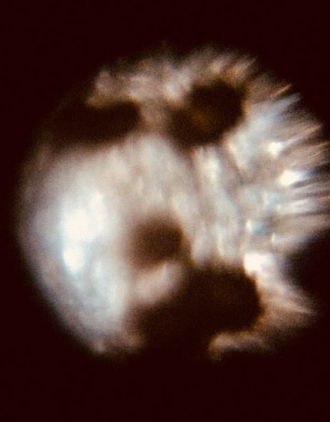
Inner wet layer with fungus It seems like there are nuclei in only some of the cells, hyphae of the fungus are winding their way through the cells and there are black spores. Later trying to figure out the criss-crossing pattern of cells we thought perhaps the peel had got folded on itself.
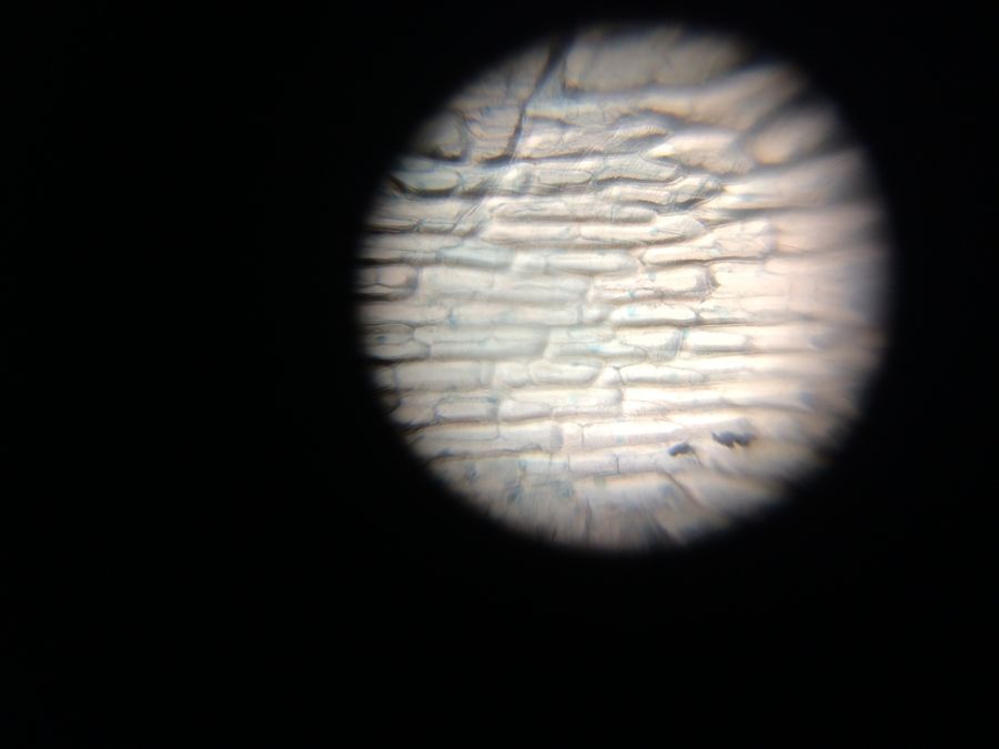
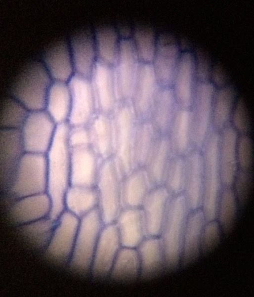
Further inside the onion the layers looked free of fungus and we saw nice cells with clear nuclei.
Further inside the onion the layers looked free of fungus and we saw nice cells with clear nuclei.
View in Media Gallery
Inner wet layer without fungus This one has two layers with a 3D effect. Seems like only the lower nuclei got stained.
View in Media Gallery
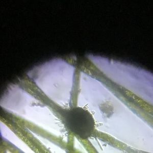
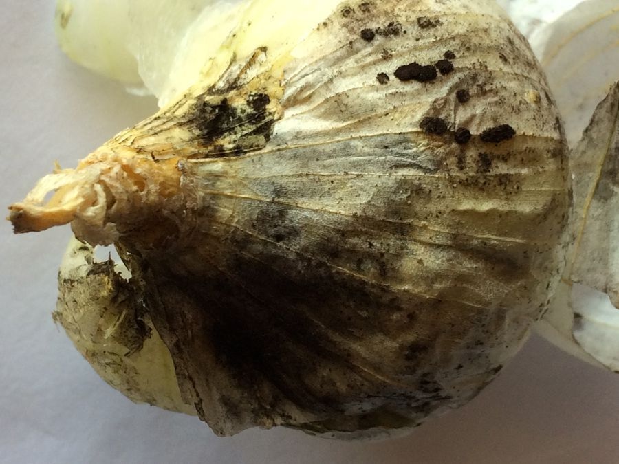
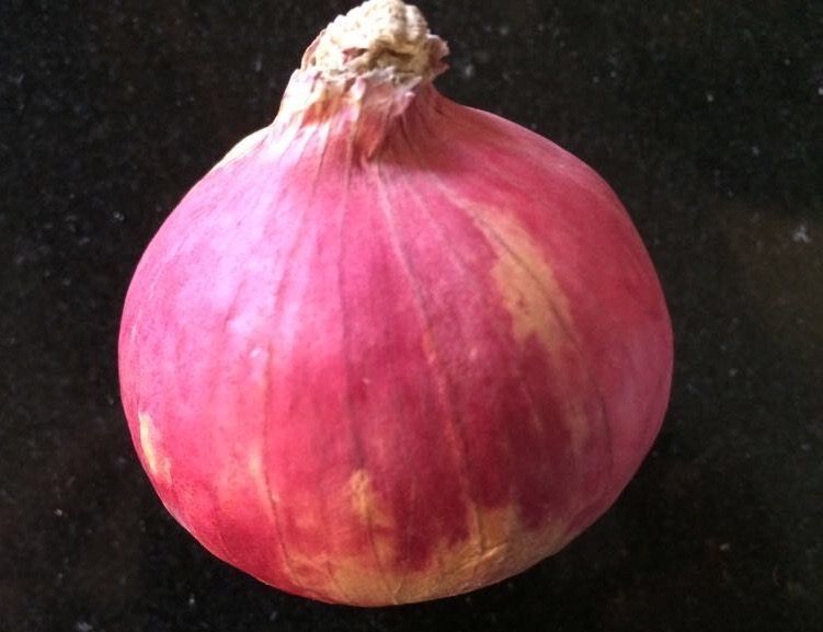
Two inner layers The next time we got a red onion. From the outside it had yellowish patches but inside, sure enough was the fungus.
View in Media Gallery
Red patchy onion
View in Media Gallery
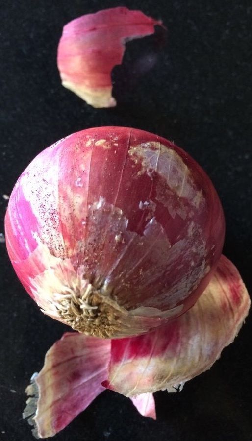
Outer skin peeled to show the fungus We looked at this dry fungus-covered skin, without staining.
View in Media Gallery
Fungus on dry skin of onion Here is an inner wet layer without fungus. This time we used a different, aqueous methylene blue which had undissolved bits that we washed off. It did not work so well. We assume that there are nuclei here but they did not get stained.
View in Media Gallery
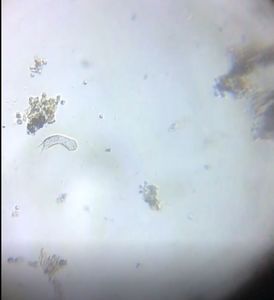
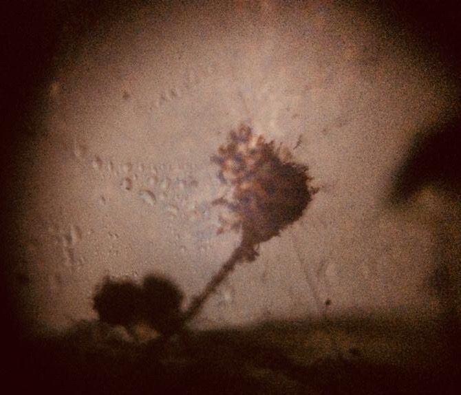
Inner wet layer without fungus At the edge of the dried skin the fungus was clearer to see.
View in Media Gallery
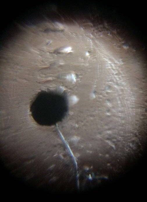
Conidiophore on the edge of the dry onion skin This one is called a “conidiophore”. The hypha (filament) is translucent and segmented. At its tip are the “conidia” or a bunch of spores. Our best guess from searching on the web is that this is Aspergillus niger , which colonises various parts of over a hundred-odd food plants, from apples to maize .
We put a drop of water on one conidiophore and its globular tip burst! Here it is, releasing its conidia into the water.
We put a drop of water on one conidiophore and its globular tip burst! Here it is, releasing its conidia into the water.
View in Media Gallery
Spores eject out under water Here’s a video of what
Aspergillus niger
can do to its host onion over just 22 days .
In Indian kitchens we deal with A. niger on a daily basis. We chuck out the infected layers of onion or wash off the powdery fungus. But this close contact with the fungus got us a bit tickly in the nose and throat. Though generally not dangerous, A. niger does contain toxins which could affect you in a variety of ways . We must take care if we do this with school students, especially in low resource environments where gloves and masks are not available.
K. Ashalatha
Jayashree Ramadas
Sign in to commentNobody has commented yet... Share your thoughts with the author and start the discussion!

 0 Applause
 0 Comments