Flowers through the Looking Glass
 Mar 06, 2018 • 10:41 PM UTC
 Unknown Location
 140x Magnification
 Microorganisms
Lara Maassen
Learn about the author...
2posts
0comments
1locations

View in Media Gallery
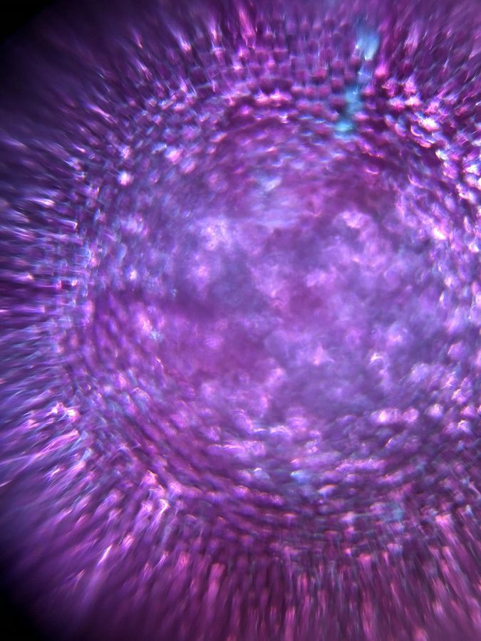
After the amazing results I obtained from my first foldscoping experience, I wanted to extend my idea and look at different flowers. I though to myself: “How will the structure from the rose I initially observed differ from other flowers and what other observations can I make about the microscopic image of flower interiors ?”
View in Media Gallery
Again, my dear Professors provided flower samples in class which I used for this experiment. I looked at three different flowers and in hindsight I wish I had taken pictures of them before using them for the samples so I could identify them now. The method I used to prepare the slides is identical to the one I used in the last post.
View in Media Gallery
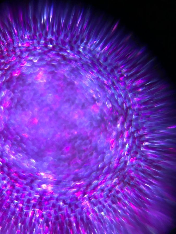
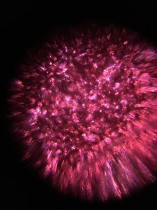
The results I obtained from this experiment were even more stunning than the rose petal. In the last two samples I observed extremely vivid, almost glowing colors, and I am unsure if this can be accounted to natural variation or an accidental change in my light source as the LED provided in the kit has two brightness settings. The observations I made about the structure of the different flowers is that they’re seems to be no very significant variation (all have tightly packed cells) but interestingly enough the pink flower does seem to have some sort of vain like structure in the bottom left corner. Any thoughts on function?
View in Media Gallery
Overall the Foldscope has served as an amazing tool – a sort of Looking Glass – into the world of flower interiors and has highlighted the beauty of the biological structures that evolution has helped create.
Sign in to commentNobody has commented yet... Share your thoughts with the author and start the discussion!
 0 Applause
 0 Comments