Spider mites on my houseplants
 Mar 22, 2015 • 11:30 PM UTC
 Unknown Location
 140x Magnification
 Microorganisms
Laks Iyer
Human observer of life. https://sukshmadarshin.wordpress.com
97posts
1255comments
5locations
View in Media Gallery
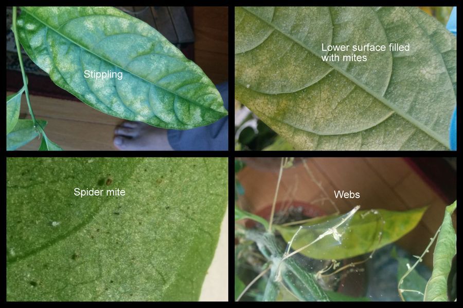
I have a Cestrum nocturnum (Raat ki Rani in hindi) plant whose fragrant flowers I greatly prize. The North-east US winters, which seems to be never-ending this year, are when I get it indoors. Recently, the plant looked very sick and started dropping leaves rapidly. On close inspection the leaves showed a stippling, as though someone threw bleach on it, and had webs with several spider mites on it. I looked under the leaf and they were there in plenty (Figure 1). I panicked, for I have many tropical plants and these can aggressively spread between plants, but before I did anything I thought I’d foldscope them.
View in Media Gallery
Figure 1
I had to do a few trials and I tried the following: 1) Directly taking some web onto a slide. 2) suspending them in water. 3) Cutting a leaf surface for direct foldscoping.
Web onto slide.
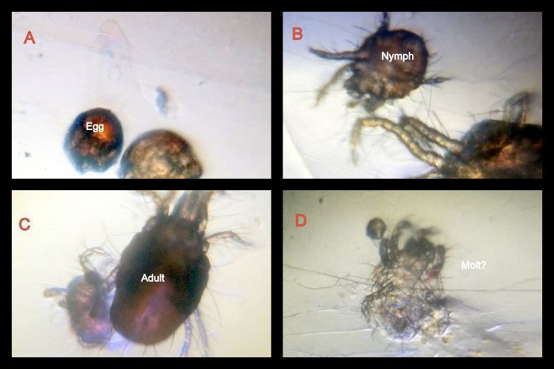
The mites were not very transparent and adults showed a reddish color. There were two marks on the dorsal side from which I infer that this is the common two-spotted spider mite Tetranychus urticae (Experts please help). Plant mites show four stages in its life-cycle, the egg, larva, Protonymph, Deutonymph and Adult. I could see eggs, and perhaps some nymphs, and an adult for sure. I am no expert on the nymphal stages, but I am sure some of them were nymphs, as they were smaller and had 8 legs, although males could also be smaller In the larval stage, the mite has 6 legs instead of the usual 8 and hence these are easy to distinguish.
I had to do a few trials and I tried the following: 1) Directly taking some web onto a slide. 2) suspending them in water. 3) Cutting a leaf surface for direct foldscoping.
Web onto slide.
The mites were not very transparent and adults showed a reddish color. There were two marks on the dorsal side from which I infer that this is the common two-spotted spider mite Tetranychus urticae (Experts please help). Plant mites show four stages in its life-cycle, the egg, larva, Protonymph, Deutonymph and Adult. I could see eggs, and perhaps some nymphs, and an adult for sure. I am no expert on the nymphal stages, but I am sure some of them were nymphs, as they were smaller and had 8 legs, although males could also be smaller In the larval stage, the mite has 6 legs instead of the usual 8 and hence these are easy to distinguish.
View in Media Gallery
Figure 2
A movie of the same follows (Figure 3). Towards the end, you can see the Chelicerae of the spider-mite mouth. Now that I see the movie again, I am wondering if the smaller mite is actually a male (and not a nymph) and what we see here is a mating act. I asked my expert friend about this, and he agrees. This video excites me to no end.
A movie of the same follows (Figure 3). Towards the end, you can see the Chelicerae of the spider-mite mouth. Now that I see the movie again, I am wondering if the smaller mite is actually a male (and not a nymph) and what we see here is a mating act. I asked my expert friend about this, and he agrees. This video excites me to no end.
Figure 3
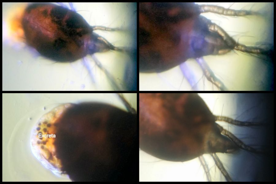
One annoying aspect was that several mites in this sample and in the ones suspended in water released the contents of their gut probably upon being pressed by the coverslip. This goop looked waxy and would inundate the view. It often contained dark spots.
One annoying aspect was that several mites in this sample and in the ones suspended in water released the contents of their gut probably upon being pressed by the coverslip. This goop looked waxy and would inundate the view. It often contained dark spots.
View in Media Gallery
Figure 4
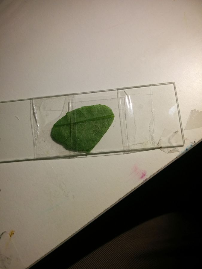
This goopy problem was worse with the spider mite in water, and so I am not posting any views of the same. The best views were of the spider mite on the leaf itself. The uneven surface of the leaf ensured that the coverslip did not crush the mite and I got some really nifty views.
This goopy problem was worse with the spider mite in water, and so I am not posting any views of the same. The best views were of the spider mite on the leaf itself. The uneven surface of the leaf ensured that the coverslip did not crush the mite and I got some really nifty views.
View in Media Gallery
Figure5
Here is one with the spider-mite feeding. You can see the gut content move. The video was taken at 2x. Spider mites pierce the leaf epidermis and feed on the mesophyll cells.
Here is one with the spider-mite feeding. You can see the gut content move. The video was taken at 2x. Spider mites pierce the leaf epidermis and feed on the mesophyll cells.
Figure 5 (Video taken at 2x)
The best thing I like about the below video is that lateral views. You can see eggs in some fields. I also feel that these have eye-spots in front of those two large marks on the dorsal surface, although they could just be the pigment extending. Many acarid spider mites are eyeless.
The best thing I like about the below video is that lateral views. You can see eggs in some fields. I also feel that these have eye-spots in front of those two large marks on the dorsal surface, although they could just be the pigment extending. Many acarid spider mites are eyeless.
Figure 6
Very briefly, spider mites are Arachnids of the acari family. Unlike insects they only have two major segments, a cephalothorax (head-thorax) and an abdomen. They have no antennae, and 8 legs (The larval stage has 6). An interesting aspect of their life is that males are haploid and females diploid. Such states often lead to cooperation and I suspect there is some kind of kin selection going on in these mites. The webbing is used to protect the mites from predators and holding onto the leaf surfaces. Some mites I have seen in the wild are really colorful. They are also trivial to find.
Now I better get that insecticidal soap out before I grieve on my favorite plant.
Sign in to commentNobody has commented yet... Share your thoughts with the author and start the discussion!

 0 Applause
 0 Comments
_300x300.jpeg)


