Imaging Tetrahymena thermophila cilia
 May 06, 2018 • 4:15 AM UTC
 Unknown Location
 140x Magnification
 Microorganisms
Laks Iyer
Human observer of life. https://sukshmadarshin.wordpress.com
97posts
1255comments
5locations
View in Media Gallery
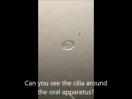
Tetrahymena thermophila are covered by about 21 rows cilia each with about 30 cilia. These 600 odd cilia allow it to propagate by beating in a coordinated way. The cilia also surround the oral apparatus to sweep food into it. These cilia are probably in the 1 micrometer range and hence imaging them requires one to use techniques like dark field and angular illumination with the foldscope. So here is a video of the same. The subcellular organelles, including the macronucleus, the micronucleus and the vacuoles are also really clear in this view. One of the most interesting subcellular organelle is the contractile vacuole, a kind of osmolarity regulator that pumps out water from within the cell. The frequency of contractions is a function of the osmolarity differential between the cell and the surrounding medium. Towards the end of the video (1:42) you see the background turning red due to a technique called Rheinberg illumination that I shall reserve for a separate post.
The assett program lists several wonderful modules that one can do with Tetrahymena and its free!! Peruse them and if possible and ask your local school science programs to get involved.
Osmolarity
The assett program lists several wonderful modules that one can do with Tetrahymena and its free!! Peruse them and if possible and ask your local school science programs to get involved.
Osmolarity
Sign in to commentNobody has commented yet... Share your thoughts with the author and start the discussion!

 0 Applause
 0 Comments
_300x300.jpeg)


