Human feces, Africa (ID: 15)
 May 25, 2018 • 8:11 AM UTC
 Unknown Location
 140x Magnification
 Unknown
Erica Schwarz
Learn about the author...
1posts
0comments
1locations

View in Media Gallery
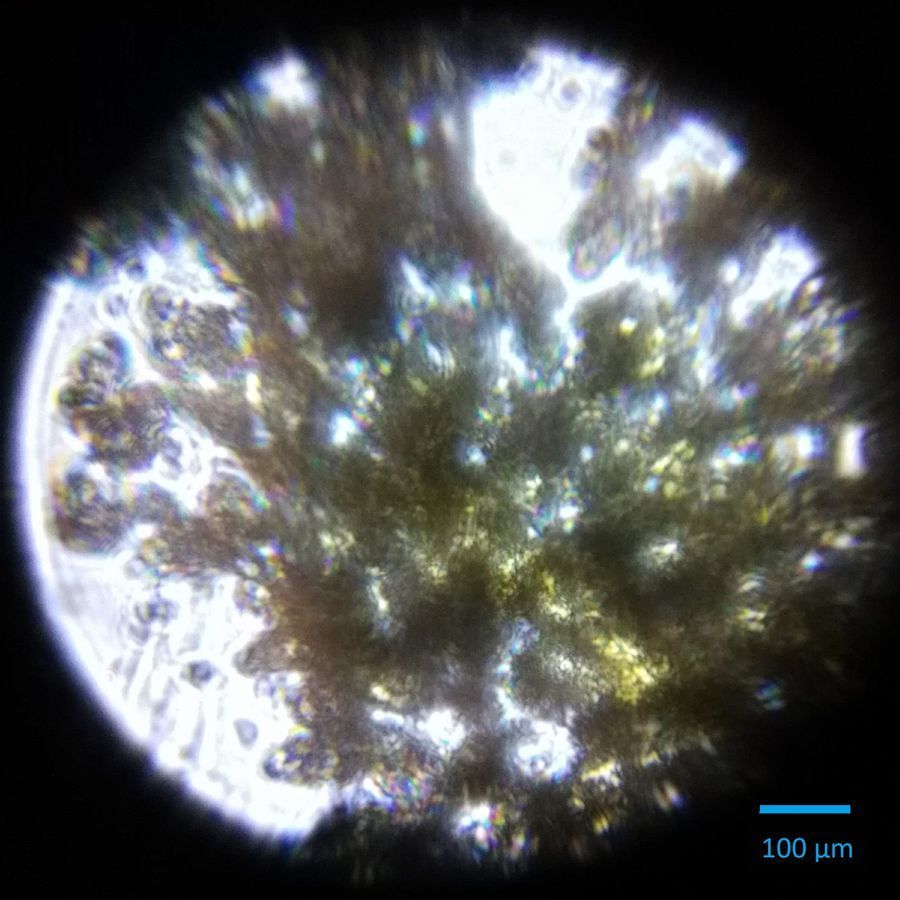
This sample consisted of human fecal matter collected from the African continent. It was imaged on a prepared slide with the foldscope using a Huawei 5X smartphone. The fecal matter smear tended to appear in clumps, resulting in some trouble finding individual cells to examine.
View in Media Gallery
However, by examining the organisms on the edges of the clumps it became a little bit easier to identify silhouettes. One in particular was recognizable as a schistosoma cercaria.
View in Media Gallery
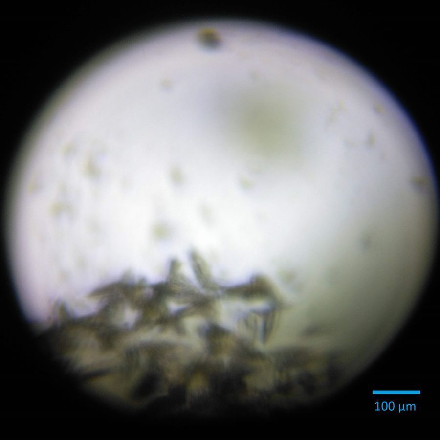
Despite some difficulty getting perfect focus in this sample, the distinct body and tail sections can be recognized. Given the geographic location of where the sample was collected, this could be identified as a schistosoma mansoni .
The scale bar was added by using the foldscope to image a measuring tape that had milliliter increments and then dividing the space between the imaged milliliter lines into ten sections to obtain the 100µm scale bar. I was a bit worried about using this method at first since I wasn’t sure about the microscopic accuracy of a macroscopic measuring tool, but the expected length for a schistosoma mansoni cercaria with tail and body as approximately 200µm which is accurately reflected by the calculated scale bar. This provided additional confidence in the accuracy of this method.
The scale bar was added by using the foldscope to image a measuring tape that had milliliter increments and then dividing the space between the imaged milliliter lines into ten sections to obtain the 100µm scale bar. I was a bit worried about using this method at first since I wasn’t sure about the microscopic accuracy of a macroscopic measuring tool, but the expected length for a schistosoma mansoni cercaria with tail and body as approximately 200µm which is accurately reflected by the calculated scale bar. This provided additional confidence in the accuracy of this method.
Sign in to commentNobody has commented yet... Share your thoughts with the author and start the discussion!
More Posts from Erica Schwarz
No more posts from this author.