Madagascar: Human Tissue Sample ID10 #BioE301C
 May 25, 2018 • 1:19 PM UTC
 Unknown Location
 140x Magnification
 Microorganisms
L Wan
Learn about the author...
1posts
0comments
1locations
View in Media Gallery
When one adventures abroad, you might have misfortune of encountering new diseases that make you their new home. As chilling as that may be, were that to occur to you, you would want to find out what would be causing you to feel unwell. Though your symptoms may vary, the main question is how to diagnose the disease and find the appropriate treatment. Depending on your context, the amount of resources and tools you have at your disposal would vary; in a low resource setting, you would have limited knowledge and fewer tools. We were presented with this challenge – given a slide with limited information, can you diagnose the disease using a Foldscope ?
Materials Foldscope Pre-stained slide Camera (on phone) Methods Initial observations were made to understand more about the sample received. Features like color, shape, and texture were observed through the Foldscope and captured using the camera. The scale was measured using the ruler on the Foldscope and cross-referenced with the sample images.
From what was observed, a guess on what kind of disease was made, and a further dive into online resources allowed us to solidify what disease was present.
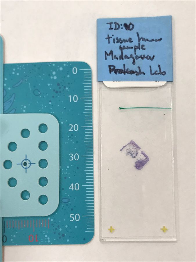
The Sample Initially we received a Foldscope and a pre-stained slide with a label describing the sample type and disease location. The slide in question was a human tissue sample with a disease originating from Madagascar.
Materials Foldscope Pre-stained slide Camera (on phone) Methods Initial observations were made to understand more about the sample received. Features like color, shape, and texture were observed through the Foldscope and captured using the camera. The scale was measured using the ruler on the Foldscope and cross-referenced with the sample images.
From what was observed, a guess on what kind of disease was made, and a further dive into online resources allowed us to solidify what disease was present.
The Sample Initially we received a Foldscope and a pre-stained slide with a label describing the sample type and disease location. The slide in question was a human tissue sample with a disease originating from Madagascar.
View in Media Gallery
Results Once the slide was inserted into the Foldscope, many different structures could be seen, but how would you find what you were looking for? Many features of varying shapes, colors, and textures could be seen, ranging from rounder cells to twisted structures.
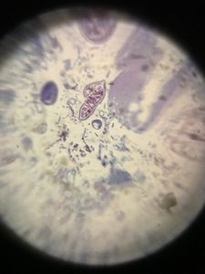
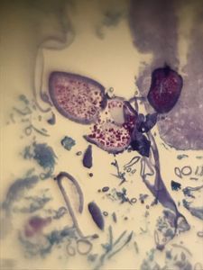
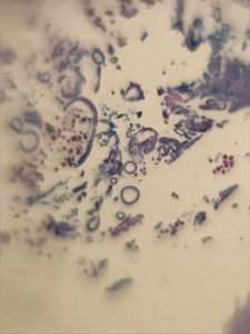
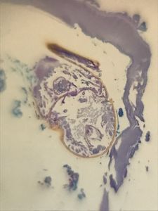
From a first glance, many structures ranging from blues to purple could be seen in the slide. Some shapes appeared as rounder structures with smaller dots within, and others as longer strands. Most structures shared features everything present, so the last image containing a brown, round object seemed promising.
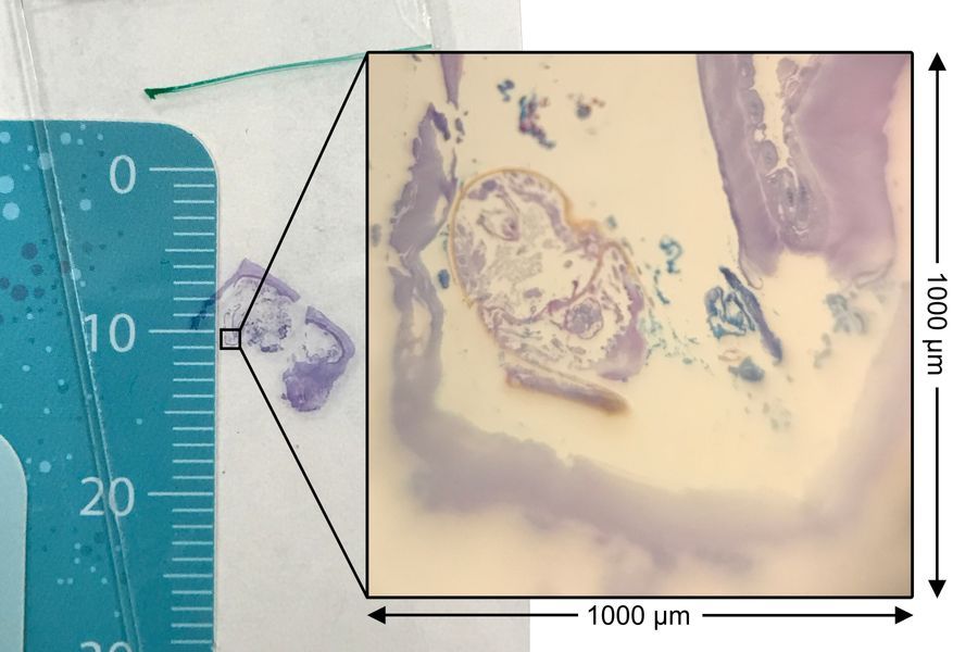
To understand the scale of the last structure, the features were compared to the Foldscope ruler. The overall shapes seen without magnification could be compared to the up close view, so the scale could be better seen, as shown below. The square shown covered one-tenth of the centimeter, so the boundary for the square covered 1 mm or 1000 microns.
To understand the scale of the last structure, the features were compared to the Foldscope ruler. The overall shapes seen without magnification could be compared to the up close view, so the scale could be better seen, as shown below. The square shown covered one-tenth of the centimeter, so the boundary for the square covered 1 mm or 1000 microns.
View in Media Gallery
With the scale analysis, the brown object in question appeared to be about 500 microns long and 200 microns wide.
Results Considering the size of the object, tissue sample type, and country of origin, the disease appeared to be Tungiasis. The mysterious brown object would then be a cross-section of a female Tunga penetrans , a flea found in tropical parts of Africa, including Madagascar. The size of our sample fit in the scale of other Tunga penetrans , which can be about 1000 microns in size.
Likely the patient who hosted this flea would have had common symptoms of a white lesion with a black dot near the skin. While recovery without intervention would have been possible with the short lifespan (< 2 weeks) of the parasite, this tissue sample would have been recovered during surgical removal, before being prepared as a slide.
Discussion Using the tools of a Foldscope, camera, and slide (with appropriate preparation tools like stains), a disease could be diagnosed with some knowledge of local diseases. While our knowledge was limited (and thus supplemented by an online search), perhaps a local health worker could be trained and equipped to better assess disease in a low-resource location.
Hoping the patient who was infected had a smooth recovery!
Sources/Further Reading Prakash Lab (http://web.stanford.edu/group/prakash-lab/cgi-bin/labsite/) https://www.dermnetnz.org/topics/tungiasis/ Edit : Looks like a good diagnosis! Oh, what’s that growing inside my foot. Story of Tunga penetrans
Sign in to commentNobody has commented yet... Share your thoughts with the author and start the discussion!
More Posts from L Wan
No more posts from this author.