African bear (ID 9) #BioE 301C
 May 27, 2018 • 8:02 PM UTC
 Unknown Location
 140x Magnification
 Unknown
Elle Robinson
Learn about the author...
3posts
0comments
1locations
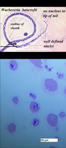
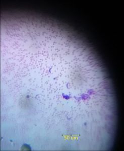
Filarial worm infection using microscope and Foldscope. A: Wucheria bancrofti a microscope with a 20 µm scale-bar. Fig. 1A is retrieved from: https://i.pinimg.com/originals/8f/d9/84/8fd9842243449728014da1bee44ed218.jpg. B: A filarial worm infection from an African bear with a 50 µm scale-bar. This image is a filarial worm infection from a African bear. Filariasis is a parasitic disease from Filarioidea roundworms spread by infected vector mosquitoes or black flies and has an associated elephantiasis symptom. It is typically characterized by the presence of these roundworms in the blood of its hosts. The roundworms are distinguishable under microscopy by its curved body and tapered tail Filariasis is diagnosed through blood film smears via the finger prick test and is found in cattle, horses, dogs, and bears. Fig. 1A shows what would be seen in a typical microscope in contrast to our device ( Fig. 1B ). Similar to the image from the microscope, the sheath can be identified in the Foldscope. The nuclei and the sheath appear more clustered, but this may be due to a larger scale in comparison, or we might be examining a different stage of development of the filiarial parasite, which appears as blue punctate in the sample. The scale-bar in Fig. 1A is 20 µm, in contrast to 50 µm for Fig. 1B. In Fig. 1B, we used a 50um scale bar to approximate the size of the parasite and the cells surrounding them. Examining a wider view of the blood smear, it becomes clear that the worm infection is prevalent in the blood since multiple punctae are visible among blood cells.
Sign in to commentNobody has commented yet... Share your thoughts with the author and start the discussion!

 0 Applause
 0 Comments