A mouth ulcer made me curious
 May 28, 2018 • 3:52 AM UTC
 Unknown Location
 140x Magnification
 Microorganisms
HARDI PARMAR
Learn about the author...
1posts
0comments
1locations
View in Media Gallery
I am Hardi Parmar, a summer visiting student with TIFR Hyderabad’s science education and outreach program ( microcosmos link ). That’s how I got my new foldscope.
My first bright idea for the foldscope was kindled by the burning sensation of an ulcer in my mouth. The ulcer must have lots of bacteria I thought, and got curious to see some. I rubbed my finger on the ulcer and its surrounding region. I suppose my fingers were fairly clean, so more stuff got transferred from the ulcer to my finger than the other way around.
Anyway, I spread the dripping liquid on a glass slide.This is how the white part of the liquid looked. I guessed it could be pus from the ulcer.

View in Media Gallery
Picture 1: White stuff around my mouth ulcer The ulcer itself was whitish. From the internet I found out that it may contain dead cells and food debris.
Three days later my ulcer had healed. During this time the stuff on the slide had mostly dried up, but a few tiny patches of saliva remained under the cover slip (which I had sealed with cello tape). Through the foldscope I looked at these patches.
Three days later my ulcer had healed. During this time the stuff on the slide had mostly dried up, but a few tiny patches of saliva remained under the cover slip (which I had sealed with cello tape). Through the foldscope I looked at these patches.

View in Media Gallery
Picture 2: Edge of cello tape with drops of saliva after 3 days

View in Media Gallery
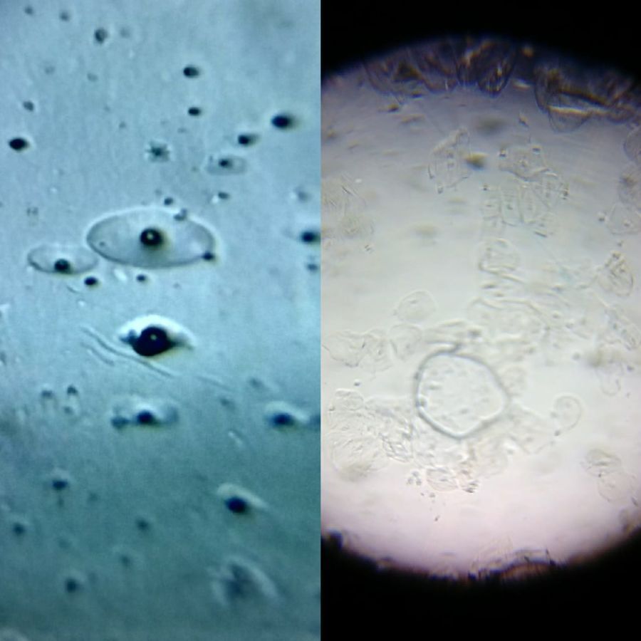
Picure 3: Patch of saliva remaining after evaporation Next are two pictures of the dried-up part. I am not sure if I see cells, or the beginnings of bacterial colonies, or something else. A biologist told me told me that the outlines of these shapes are too smooth and rounded for them to be cheek cells. (See more such shapes at the end of this post.)

View in Media Gallery
Picture 4: Dried-up saliva in LED light

View in Media Gallery
Picture 5: Dried-up saliva in sunlight The bluish pictures are taken with LED light and the grey ones in sunlight.
I also got to see some other things, like..
I also got to see some other things, like..


These may be what they call irregular or filamentous bacterial colonies .
On the internet I also found this amazing visualisation of communities of mouth bacteria .
A few days later I took a sample of my cheek cells. Here is a video of the sample:
Video 1: My cheek cells
The cheek cells are irregular in shape. A couple of smooth oval shapes are also seen, at 7 seconds and 15-18 seconds. They look very much like the shapes that I saw in the previous sample after drying (Pictures 4 and 5). I wonder if they are cells or not.

View in Media Gallery
Picture 9: Oval shape cell with cheek cell There seem to be some tiny moving things around the cells. Perhaps they are motile bacteria?
– Hardi Parmar
– Hardi Parmar
Sign in to commentNobody has commented yet... Share your thoughts with the author and start the discussion!
More Posts from HARDI PARMAR
No more posts from this author.