A Lousy Story
 May 29, 2018 • 4:06 AM UTC
 Unknown Location
 140x Magnification
 Microorganisms
Bhavya sahithi velagapudi
Learn about the author...
1posts
1comments
1locations

View in Media Gallery
I am Bhavya Sahithi a summer visiting student with TIFR Hyderabad’s science education and outreach program ( microcosmos link ). That’s how I got my new foldscope.
I managed to find some head lice and made quite a few observations on them — their eggs, the male-female differences and the feces of this fascinating parasitic insect.
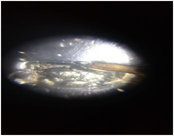
EGG OF LOUSE
Here is the egg of a head louse observed under the foldscope.
I managed to find some head lice and made quite a few observations on them — their eggs, the male-female differences and the feces of this fascinating parasitic insect.
EGG OF LOUSE
Here is the egg of a head louse observed under the foldscope.
View in Media Gallery
Egg of a head louse The video pans over the whole egg showing the transparent shell and the baby louse inside it. Attached to the egg are some wiry structures. I don’t know what they are.
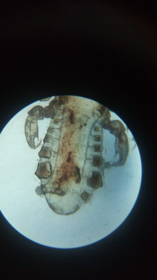
STRUCTURE OF LIVE MALE LOUSE
Here is a live louse. Figures 2 and 3 show its head and the abdomen. The head has a pair of antennae. Below that we see its three pairs of legs. The abdomen has 7 segments. There is brownish-red blood in the abdomen.
STRUCTURE OF LIVE MALE LOUSE
Here is a live louse. Figures 2 and 3 show its head and the abdomen. The head has a pair of antennae. Below that we see its three pairs of legs. The abdomen has 7 segments. There is brownish-red blood in the abdomen.
View in Media Gallery
Head of male louse

View in Media Gallery
Abdomen of male louse We can tell that this is a male louse because it has a rounded back end (posterior). Also in the male the first pair of legs are large which helps them hold the female louse during copulation.
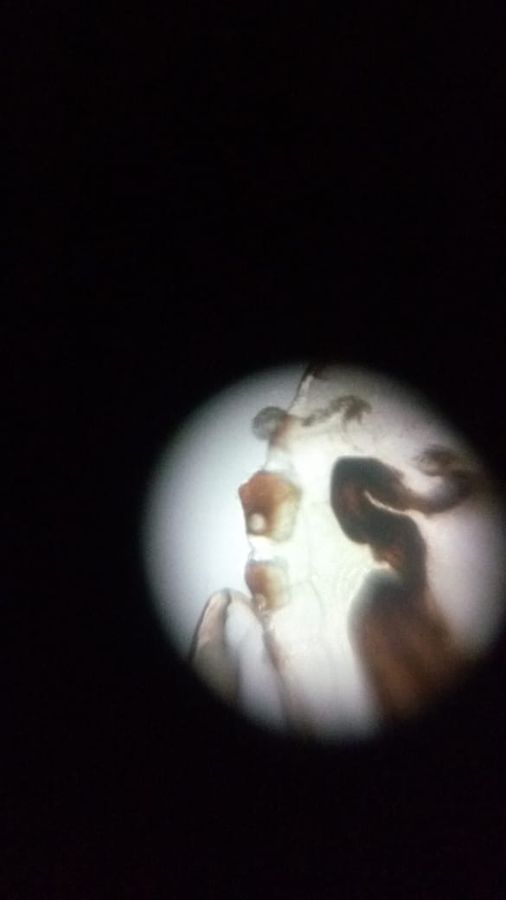
This video pans over the male louse. We can see the claws at the end of its legs which help to hold on to the hair and the tactile bristles which probably help them sense movement or danger .
OBSERVATION OF DEAD LOUSE
I pressed a male louse with my thumb nail and killed it. The next video shows how it looked through the foldscope. When I pressed the back of foldscope to focus it I found some internal liquid running through the louse (see 40 seconds onwards).
View in Media Gallery
Killed male louse abdominal part
View in Media Gallery
Fluid in abdomen of killed male louse FECES OF FEMALE LOUSE
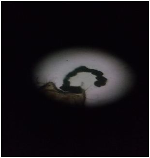
Next I observed a female louse. I know it was female because of the “W” shaped gonopods at the posterior end . Initially I saw a small dark round structure between the gonopods. Then we went for lunch. When I came back after an hour I saw this curved structure, like beads on a string, which had emerged from it.
Next I observed a female louse. I know it was female because of the “W” shaped gonopods at the posterior end . Initially I saw a small dark round structure between the gonopods. Then we went for lunch. When I came back after an hour I saw this curved structure, like beads on a string, which had emerged from it.
View in Media Gallery
Feces from female louse
I confirmed from the internet that it is the feces .
FECES OF MALE LOUSE
Later I found a male head louse with a similar dried-up beads on string type structure of feces. We see bristle-like structures at its posterior end. Although the males have a rounded back end, but this may appear pointed because the penis often protrudes through the genitals.

View in Media Gallery
Feces of male louse
View in Media Gallery
Pointed edge of male louse I learnt that the human body louse
Pediculus humanus humanus
has the smallest insect genome known .
Those lice are now exterminated, but they taught me a lot. I started thinking in a new way about pests and parasites, how we relate to them, and how they carry on their own lives among us.
– Bhavya Sahithi
Pediculus humanus humanus
has the smallest insect genome known .
Those lice are now exterminated, but they taught me a lot. I started thinking in a new way about pests and parasites, how we relate to them, and how they carry on their own lives among us.
– Bhavya Sahithi
Sign in to commentNobody has commented yet... Share your thoughts with the author and start the discussion!
More Posts from Bhavya sahithi velagapudi
No more posts from this author.