Onion outside in
 Jun 12, 2018 • 4:53 AM UTC
 Unknown Location
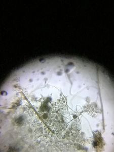
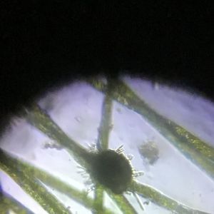
 140x Magnification
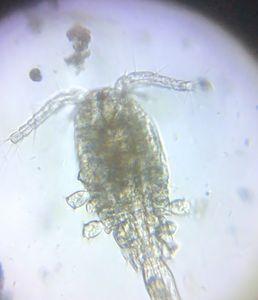
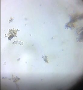
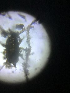
 Microorganisms
Jayashree Ramadas
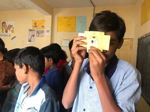
We are a group of students, volunteers and staff working with TIFR Hyderabad's Science Education and Outreach program: http://www.tifrh.res.in/~outreach/
39posts
26comments
2locations
View in Media Gallery
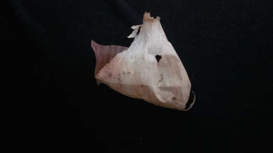
We started with an onion from the outside. First there’s a dried up layer or ‘tunic’ that we don’t eat.
View in Media Gallery
Tunic of onion
View in Media Gallery
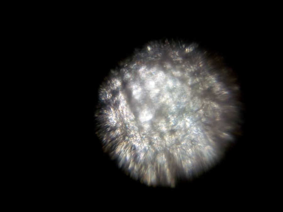
Onion tunic under foldscope The cells in the tunic must be dead. Even the cell walls are disintegrated, quite unlike the dead xylem cells that we saw in matchsticks . Later we found, in the wonderful Walter Dioni’s website, that we might also see crystals of calcium oxalate and other chemicals in the tunic of the onion . That is for other times.
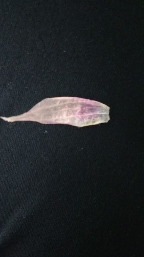
Next below it was a semi-dried layer which too we often discard.
Next below it was a semi-dried layer which too we often discard.
View in Media Gallery
Semi-dried layer of onion
View in Media Gallery
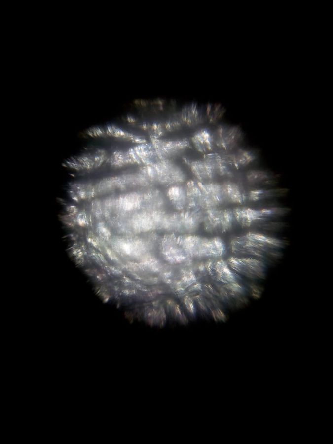
Semi-dried layer under foldscope In this layer the cell walls still remain somewhat visible.
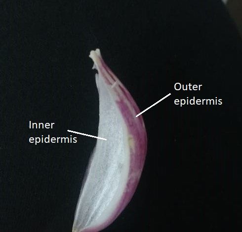
Now come the nice fleshy layers of onion, or ‘scales’. You can remove the skin, or ‘epidermis’, from the outer or the inner side of each scale. The textbook tells us to take the inner epidermis but we tried both sides, about 3 samples of each. The outer epidermis of all the scales was pinkish in color and did not stain so easily. But it did have a nice surprise for us!
Now come the nice fleshy layers of onion, or ‘scales’. You can remove the skin, or ‘epidermis’, from the outer or the inner side of each scale. The textbook tells us to take the inner epidermis but we tried both sides, about 3 samples of each. The outer epidermis of all the scales was pinkish in color and did not stain so easily. But it did have a nice surprise for us!
View in Media Gallery
Inner and outer epidermis Here is the outer epidermis of one scale:
View in Media Gallery
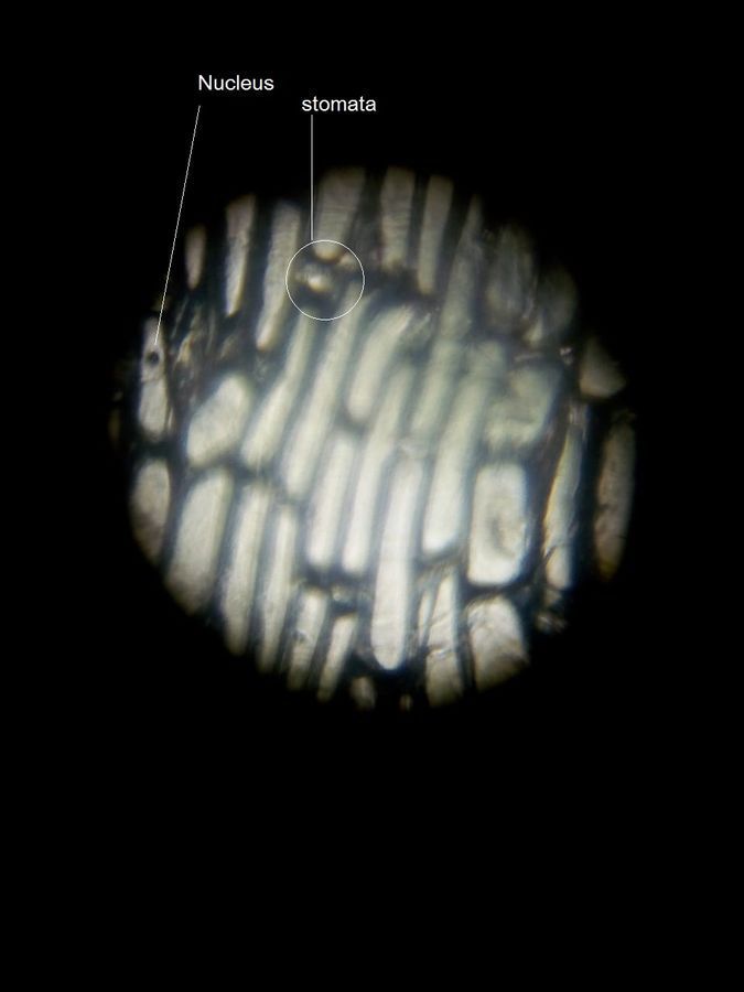
Outer epidermis of onion scale
Here is the inner epidermis:
Here is the inner epidermis:
View in Media Gallery
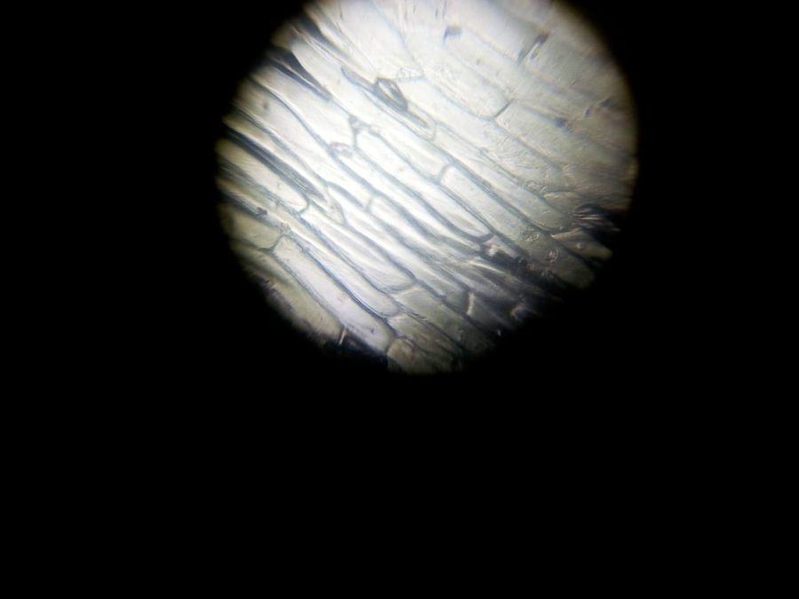
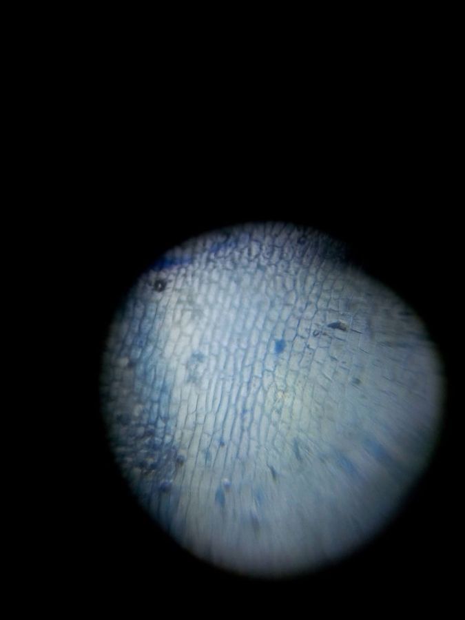
Inner epidermis of onion scale
View in Media Gallery
Inner epidermis stained with methylene blue
View in Media Gallery
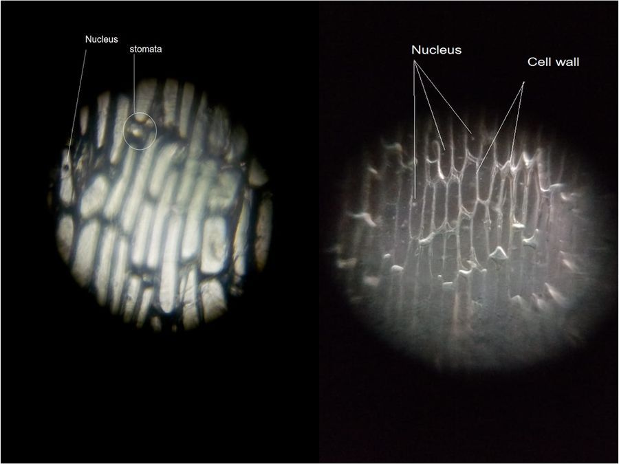
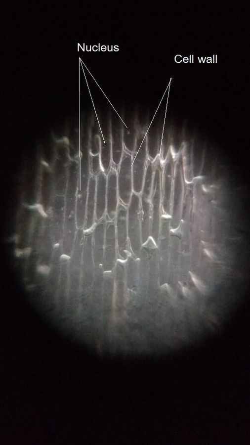
Inner epidermis with dark field The dark field image is taken with a finger in front of the camera lens held in sunlight. In it the nuclei are seen clearly, better than in the stained slide.
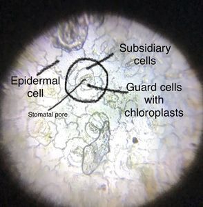
We found that the cells of the inner epidermis were fairly uniform in shape and size. In the outer epidermis the cells were of different shapes and sizes. We were further surprised to find stomata in the outer epidermis! Again, Walter Dioni told us , the scales are actually modified onion leaves. If the scale grew out into a leaf then its outer epidermis would become the lower epidermis of the leaf. That’s why the outer epidermis of each onion scale has stomata and the inner one (usually) doesn’t.
Onion cells are larger than most plant cells: the average size of onion skin cells is about 400 microns . In comparison, most other plant cells are between 10-100 microns in size .
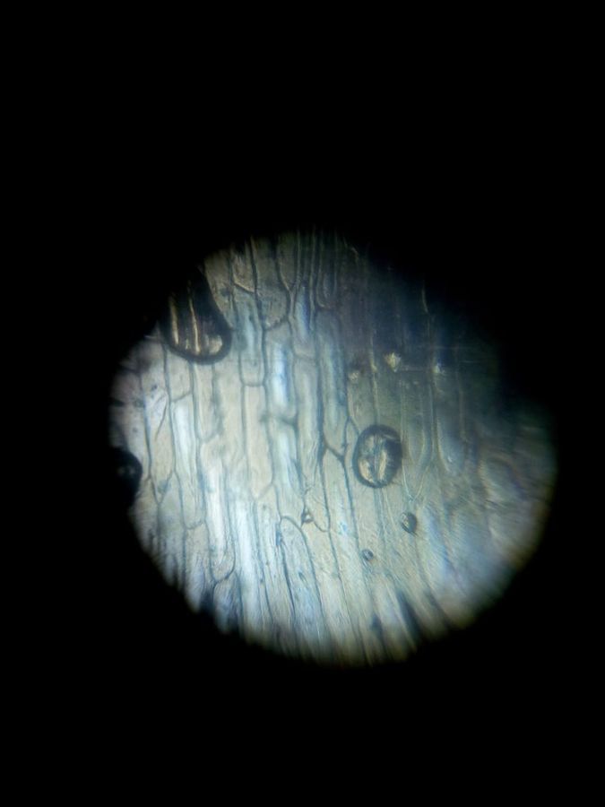
Onion is Allium cepa . We also looked at garlic, or Allium sativum . There is just one epidermis surrounding the garlic clove, which is actually a bulb. This is what it looked like.
We found that the cells of the inner epidermis were fairly uniform in shape and size. In the outer epidermis the cells were of different shapes and sizes. We were further surprised to find stomata in the outer epidermis! Again, Walter Dioni told us , the scales are actually modified onion leaves. If the scale grew out into a leaf then its outer epidermis would become the lower epidermis of the leaf. That’s why the outer epidermis of each onion scale has stomata and the inner one (usually) doesn’t.
Onion cells are larger than most plant cells: the average size of onion skin cells is about 400 microns . In comparison, most other plant cells are between 10-100 microns in size .
Onion is Allium cepa . We also looked at garlic, or Allium sativum . There is just one epidermis surrounding the garlic clove, which is actually a bulb. This is what it looked like.
View in Media Gallery
Epidermis of garlic clove Bhavya Sahithi ( Bhavya on Microcosmos )
Jayashree Ramadas
Jayashree Ramadas
Sign in to commentNobody has commented yet... Share your thoughts with the author and start the discussion!

 0 Applause
 0 Comments