Cheek and inner lip cells
 Jun 12, 2018 • 11:44 PM UTC
 Unknown Location
 140x Magnification
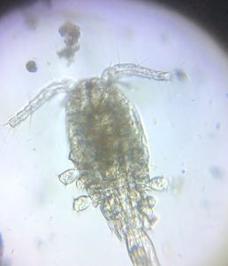
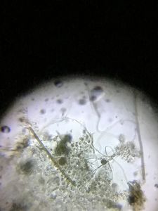
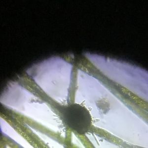
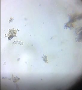
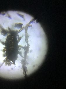
 Microorganisms
Jayashree Ramadas
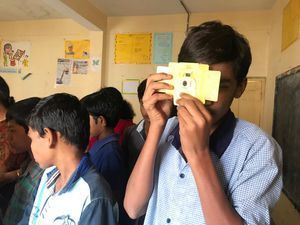
We are a group of students, volunteers and staff working with TIFR Hyderabad's Science Education and Outreach program: http://www.tifrh.res.in/~outreach/
39posts
26comments
2locations

View in Media Gallery
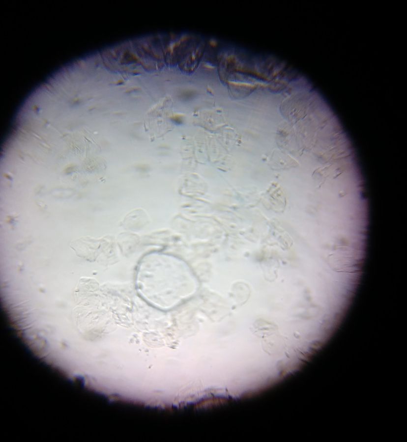
At about 60 microns in size, human cheek cells are larger than most other animal cells (which are typically 10-30 microns) . They are also easily found, by gently chewing the inside of your cheek and scraping out the surface with a spoon or a finger.
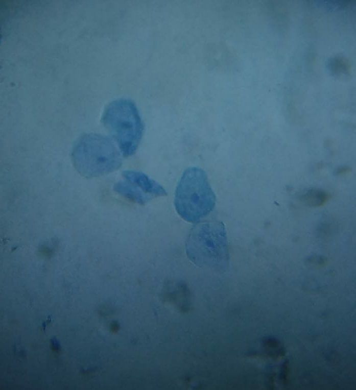
These are some images of Hardi’s cheek cells, first without stain and second stained with methylene blue.
These are some images of Hardi’s cheek cells, first without stain and second stained with methylene blue.
View in Media Gallery
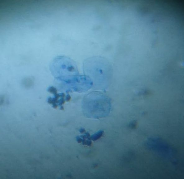
Cheek cells – without stain
View in Media Gallery
Cheek cells – with stain The dots in the cells may be organelles, or bacteria. We wondered if the larger cylindrical bodies inside the cells (labelled ‘?’) might be mitochondria.
For comparison with cheek cells Hardi thought to sample the cells from her lower inner lip. She took 4-5 samples of each kind.
For comparison with cheek cells Hardi thought to sample the cells from her lower inner lip. She took 4-5 samples of each kind.
View in Media Gallery
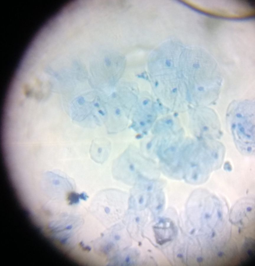
Cells of inner lip – with stain
View in Media Gallery
Cells of inner lip – with stain (2)
View in Media Gallery
Cheek cell – with stain In all the samples the cheek cells looked larger than the inner lip cells and they also had a more prominent nuclei. Anyone else like to try this?
Hardi Parmar ( Hardi on Microcosmos )
Jayashree Ramadas
Hardi Parmar ( Hardi on Microcosmos )
Jayashree Ramadas
Sign in to commentNobody has commented yet... Share your thoughts with the author and start the discussion!

 0 Applause
 0 Comments