ISOLATION OF LITTER DECOMPOSING MICROORGANISMS IN MANNAR HILL RANGE – THE WESTERN GHATS
 Jul 09, 2018 • 12:20 PM UTC
 Unknown Location
 140x Magnification
 Fungi
Fungi
Ramya Dinesh
Learn about the author...
2posts
0comments
1locations

View in Media Gallery
I am E.K. Ramya, working as a Project Assistant under the foldscope project of DBT. Our PI is Dr. S. Sharmila, Assistant Professor, Vellalar College for Women, Erode, Tamil nadu.
Litter decomposition is a process of decaying the dead organic material – with the help of microorganisms. It is one of the crucial role in the forest ecosystem. For decomposition Bacteria and Fungi play a major role. Identification of litter decomposing microorganisms in study area was the 4 th step of my study. For this study, I have collected the litter samples in litter bags during May 2018 and the collected samples are used for further microbial analysis. Analysis of microbes from litter (May 2018):
Litter sample contains mass of microorganisms. It was isolated in the culture medium using Nutrient Agar Medium.
For isolation I have followed 4 important steps:
Step 1: Bacterial Colony formation
Step 2: Isolation of bacteria in Pure culture
Step 3: Gram staining
Step 4: Biochemical test
Step 1: Bacterial Colony formation:
Bacteria grow on solid media as colonies. A colony is known as visible mass of microorganisms originating from single mother cell.
View in Media Gallery
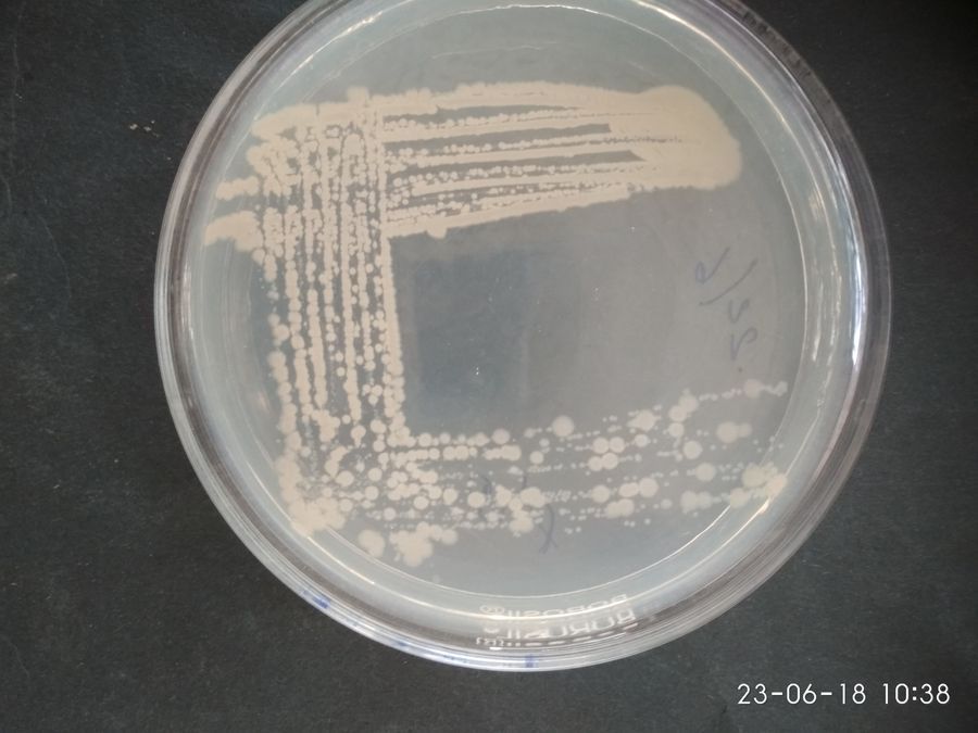
This pic… shows the Nutrient Agar Media with Beautiful Bacterial Colonies The plate showing the bacterial colonies. I have captured this picture in my microbial laboratory. The pic… also indicates numerous different colonies. The Colonies are occurring in different size & shape. I have calculated number of colonies in single plate with the help of colony counter. Step 2: Isolation of bacteria in Pure culture:
Different bacterial colonies were isolated and inoculated into other separate media. After 4 or 5 sub culturing I have maintained two pure cultures.
Different bacterial colonies were isolated and inoculated into other separate media. After 4 or 5 sub culturing I have maintained two pure cultures.
View in Media Gallery
This pic… shows the unknown bacteria in Nutrient Agar Media – Quadrant Streak plating For separation of gram positive and gram negative I have done Staining process. With the support of foldscope under high magnification I clearly viewed beautiful colour differentiation in gram staining process. Two pure cultures:
Gram positive Gram negative
Gram positive Gram negative

View in Media Gallery
Foldscope view of Rod shaped Gram-positive bacteria– (violet)

View in Media Gallery
Foldscope view of Rod shaped Gram-negative bacteria – (Pink) Biochemical test:
This is the important step for differentiate species from genera.
Biochemical Test for Gram- Positive bacteria
Indole test Methyl Red test Vioges Proskauer test Citrate utilization test Indole test (-ve)
This is the important step for differentiate species from genera.
Biochemical Test for Gram- Positive bacteria
Indole test Methyl Red test Vioges Proskauer test Citrate utilization test Indole test (-ve)

View in Media Gallery
a) No pink ring formation Methyl Red test (-ve)
View in Media Gallery
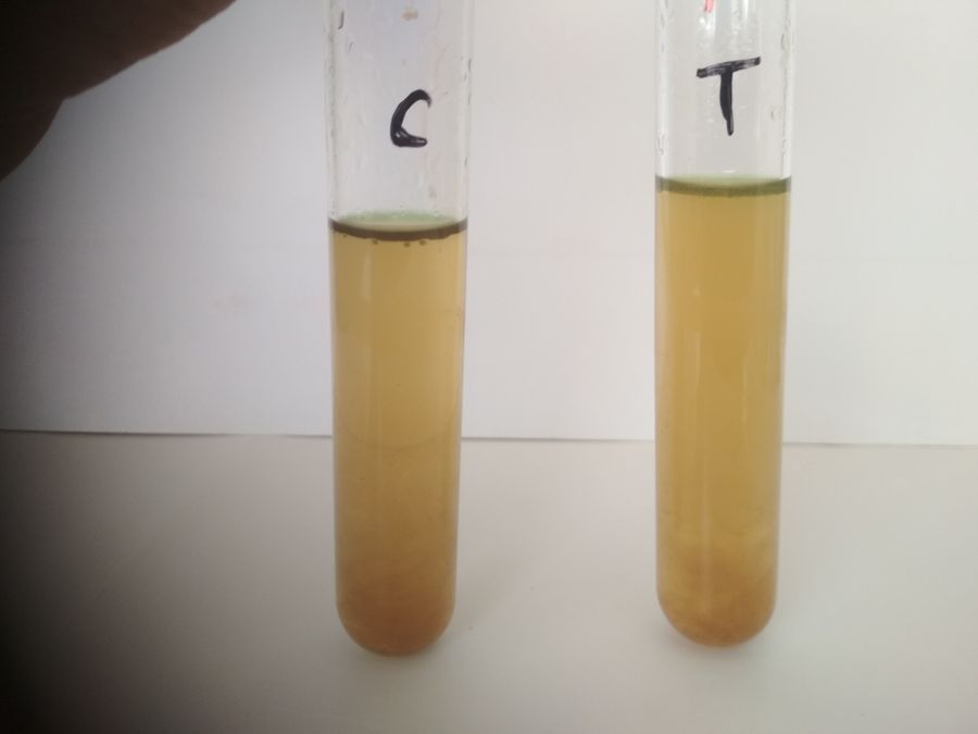
b) No colour changes (pink) Vioges Proskauer test (+ve)
View in Media Gallery
c) Pink ring formation Citrate utilization test (+ve)

View in Media Gallery
d) Colour changes (Blue) Biochemical Test for Gram- Negative bacteria Indoletest (-ve)

View in Media Gallery
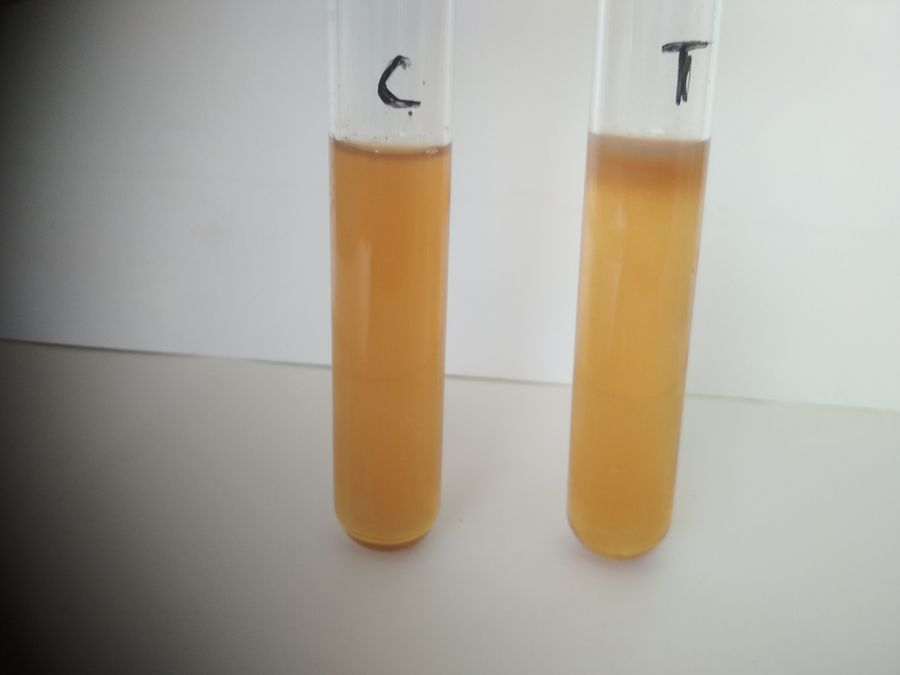
e) No pink ring formation Methyl Red test (-ve)

View in Media Gallery
f) No colour changes (pink) Vioges Proskauer test(-ve)

View in Media Gallery
g) No pink ring formation Citrate utilization test (+ve)

View in Media Gallery
h) Colour changes (Dark blue)
As per the schedule of work plan, half of the work in process. Later I will upload remaining part of the study (Isolation of micro – organisms) with the help of foldscope magnification.
As per the schedule of work plan, half of the work in process. Later I will upload remaining part of the study (Isolation of micro – organisms) with the help of foldscope magnification.
Sign in to commentNobody has commented yet... Share your thoughts with the author and start the discussion!

 0 Applause
 0 Comments