Bitten by a tick
 Apr 30, 2015 • 10:04 PM UTC
 Unknown Location
 140x Magnification
 Microorganisms
Laks Iyer
Human observer of life. https://sukshmadarshin.wordpress.com
97posts
1255comments
5locations
View in Media Gallery
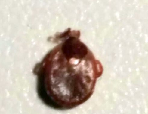
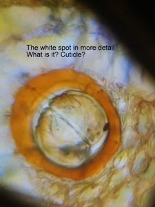
The coming of spring brings great joy to natural history lovers in the northeastern US, although there is one scourge who is also very active around this time and no one’s favorite– The tick. While ticks per se are just a minor nuisance, the diseases they transmit are quite dangerous especially if untreated. In particular the DC area has seen a rise in Lyme’s disease, caused by the bacterium Borrelia burgdoerferi, and transmitted by the Deer tick. Naturally, there is a lot of paranoia with tick bites around my regions. Recently, a member of my family was bitten by a tick. The tick, about 5mm in length, was not engorged (filled with blood), rather flat, hard-bodied and was firmly lodged in the back of the individual. I took a pair of forceps, went as close to the head as possible and pulled it out. Pulling it out wasnt as easy as I thought it would be as the tick was well lodged into the skin. A careful examination revealed that this was a female lone-star tick ( Amblyomma americanum ) that doesnt trasmit Lyme’s disease (relief), although at times it is a vector for Ehrlichiosis, tularemia and Southern tick-associated rash illness (I didnt mention these at home). The lone-star tick has a characteristic white dot/star on its back.
Note the white spot on the back
Note the white spot on the back
View in Media Gallery
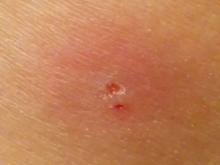
Note the two chelicerae insertion points in the wound
View in Media Gallery
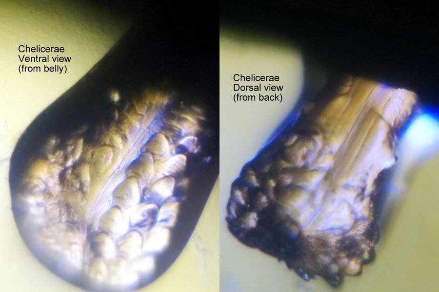
I put the tick in 70% ethanol overnight and then into a solution of lactoglycerol (50% lactic acid: 50% glycerol, both commonly available beauty products). The lactoglycerol step was an attempt to clear the tick as it was quite sclerotized and opaque (see above left). I kept it for many days in the lactoglycerol solution till I got to it in my foldscope. One of the things that I wanted to understand was the reason for it being so hard to pull the tick out, and I think I got the answer. Look at the following foldscope pictures.
Click on images for hi-res images.
Click on images for hi-res images.


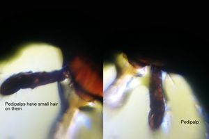

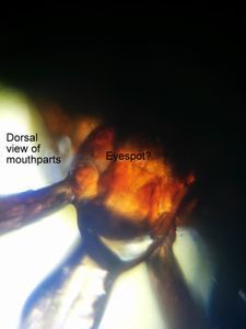
Observations.1. The mouth of the tick has three parts (look at bottom row and the middle picture of the middle row), the two outer palps, the chelicerae and a hypostome that emerges from the chelicerae. The chelicerae firmly attach into the individual with their rasping hooks on both sides as seen in the pictures above (bottom row middle). You can see that the chelicerae are symmetrical. The hypostome which also has teeth emerges between the chelicerae and is a straw like structure through which the tick enjoys its blood meal and injects saliva and gut contents, which result in transmission of pathogens. I had pulled the tick well enough to display the chelicerae. Perhaps the hypostome is withdrawn into the chelicerae, or perhaps stuck in the individual. The teeth on the chelicerae point towards the tick and hence when you pull on them, the chelicerae teeth just hold on like hooks.
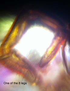
2. The ends of the legs have a hook-like structure which further hold steadfastly on the individual.
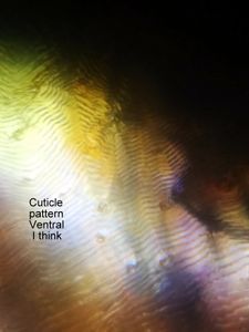
3. I found the surface cuticular patterns very interesting. It almost looks like a fingerprint. I wonder if this is variable between individual ticks.
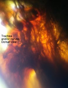
4. The clearing by lactoglycerol shows trachea within the tick. Its a good reagent, but the tick becomes very delicate after sometime and I squashed it flat inadvertantly, thereby ending my tick adventure. I might still preserve the head and try to make a permanent mount of it. Quite an adventure.
2. The ends of the legs have a hook-like structure which further hold steadfastly on the individual.
3. I found the surface cuticular patterns very interesting. It almost looks like a fingerprint. I wonder if this is variable between individual ticks.
4. The clearing by lactoglycerol shows trachea within the tick. Its a good reagent, but the tick becomes very delicate after sometime and I squashed it flat inadvertantly, thereby ending my tick adventure. I might still preserve the head and try to make a permanent mount of it. Quite an adventure.
Sign in to commentNobody has commented yet... Share your thoughts with the author and start the discussion!

 0 Applause
 0 Comments
_300x300.jpeg)


