Pollen hunter project–III- Investigating pollen size with a scale
 May 13, 2015 • 9:35 PM UTC
 Unknown Location
 140x Magnification
 Microorganisms
Laks Iyer
Human observer of life. https://sukshmadarshin.wordpress.com
97posts
1255comments
5locations
View in Media Gallery
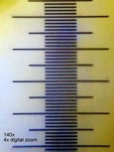
I was displaying my pollen slides to kids around my place and one of them, all of 5 years, who measures everything with a scale asked , “How small are they?”. I mumbled my way through deftly, ignoring the question but it rang for a while in my head after. Measurement is an integral part of science and this was one aspect that I had conveniently ignored through this project. Could I create a digital scale that I could use over and over. I pulled out a stage micrometer that I had bought some time back. These cost about $11 on amazon , and their smallest division is 0.01 mm or 10 microns and they have a 100 divisions in total (Figure 1A). I have a Google phone that I measured has 4x digital zoom (Figure 1B).
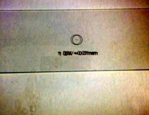
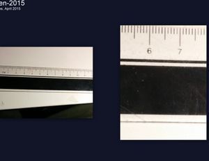
Figure 1A and 1B
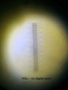
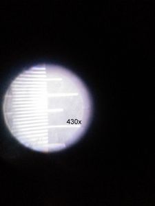
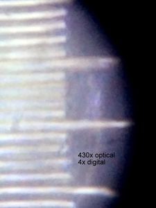
For all my pollen pictures, I mostly use the 140x optical zoom and the 4x digital zoom, or on rare occasions the 400x optical zoom with a 4x digital zoom. This mean that if I create a template picture of the stage micrometer at these optical and digital magnifications, I shall have a scale that I can superimpose (using google picasa) and get an approximate size of the pollen or any object of study. First I needed to have pictures of the slide micrometer.
Optical magnification
No Digital zoom
4x Digital zoom
140x
400x
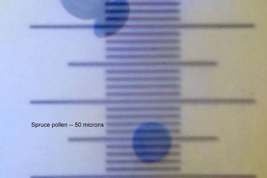
As long as I use the same digital and optical zoom on my sample and capture pictures with my Google phone, I can superimpose any picture with these scales using google picasa and measure distances knowing that each division is 10 microns. Wow this is exciting. I cant wait to wake up and tell the 5 year old that I know the size of the pollen. A few measurements of the different objects I have seen with the foldscope follow. If you see a logical error in my reasoning, let me know.
Object and calculated measurement
Image
Spruce pollen – 50 microns
Object and calculated measurement
Image
Spruce pollen – 50 microns
View in Media Gallery
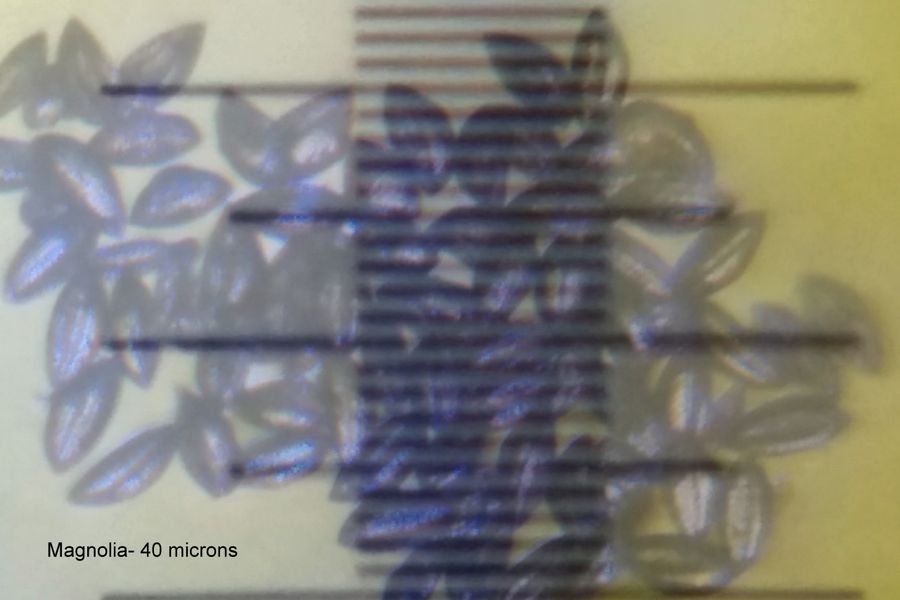
Magnolia pollen – 40 microns
View in Media Gallery
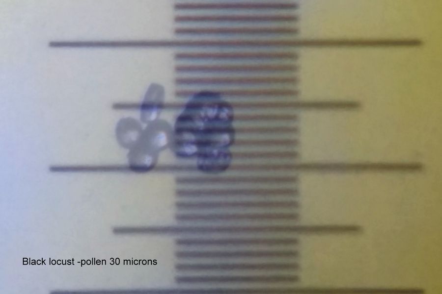
Black locust pollen- 30 microns
View in Media Gallery
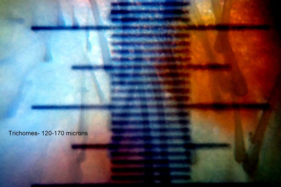
Trichomes 120-170 microns
View in Media Gallery
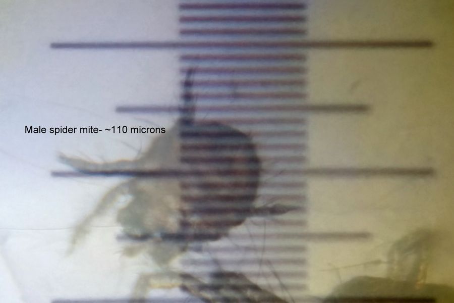
Male spider mite – ~110 microns
View in Media Gallery
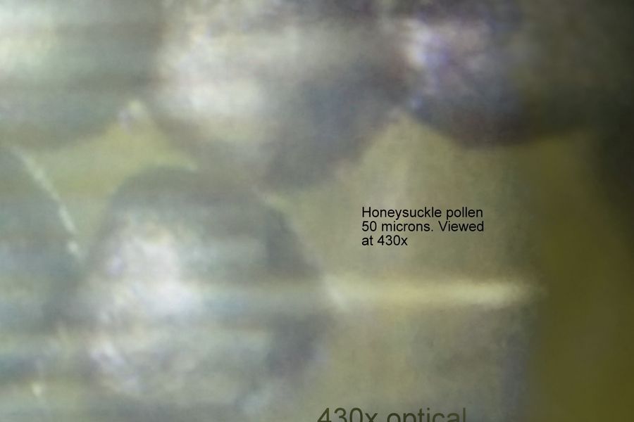
Honey-suckle pollen at 400x- 50 microns
View in Media Gallery
Sign in to commentNobody has commented yet... Share your thoughts with the author and start the discussion!

 0 Applause
 0 Comments
_300x300.jpeg)


