Posterior Spiracle of Maggots ( Musca domestica Larvae)
 Aug 29, 2018 • 3:10 AM UTC
 Unknown Location
 140x Magnification
 Microorganisms
Meignanalakshmi Sundaram
I am Dr.S.Meignanalakshmi, working as Professor, at the Directorate of Centre for Animal Health Studies, TANUVAS, Chennai-51. Working on Foldscope project on "Foldscope for diagnosis of Rumen Acidosis and parasitic infections in cattle" sanctioned by DBT
66posts
8comments
1locations
View in Media Gallery
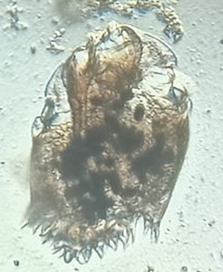
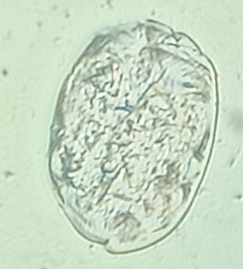
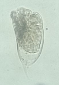
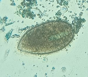
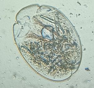
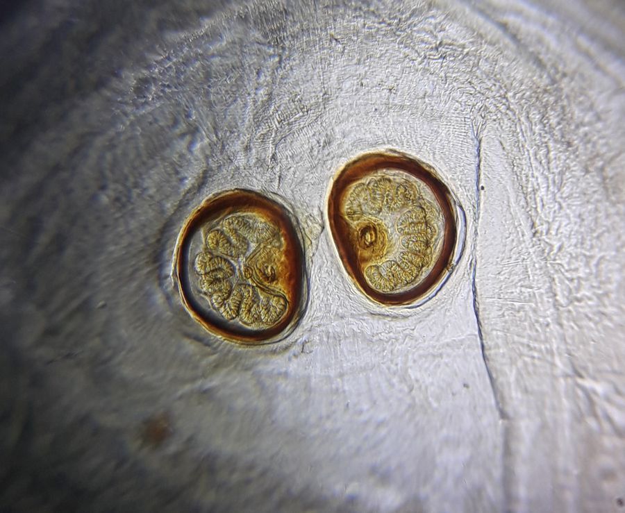
Here’s a beautiful and detailed picture of the processed Posterior Spiracle of the Musca domestica Larvae, which was collected from an infected wound.
The larvae was boiled in 10% NaOH for about 5 mins and was thoroughly rinsed in water. It was then dehydrated for 2 – 5 mins in increasing concentrations of alcohol ( 70%, 90% and 100%). The larvae was then kept in a clearing agent ( carbolic acid). The cleared larvae was mounted in DPX mountant and examined under foldscope. The posterior end of the larvae consists of
a Pair of brown ‘D’ shaped spiracles A chitinised ring (button) and 3 sinuous ‘m’ shaped stigmatic slits on each spiracle.

View in Media Gallery
Sign in to commentNobody has commented yet... Share your thoughts with the author and start the discussion!

 0 Applause
 0 Comments