Piranha of the Japurá River
 Sep 03, 2018 • 9:57 PM UTC
 Brazil
 140x Magnification
 Animals
Rebecca Konte
Scientific artist, using art as a tool for scientific storytelling. http://www.rebeccakonte.com/
27posts
25comments
22locations
View in Media Gallery
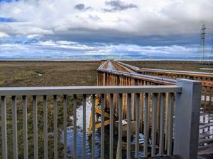
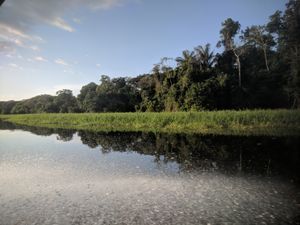
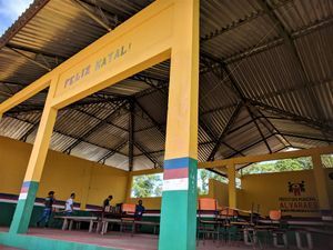
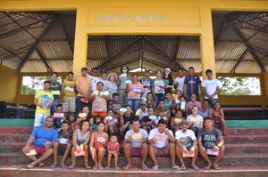
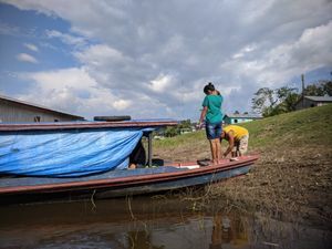
We held a workshop in a fishing community along the Japurá River.
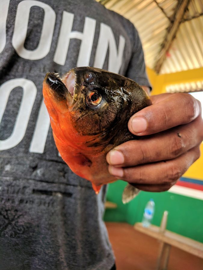
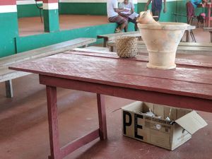
There comes a time in every workshop where our attention shifts from building the foldscope to “Now what are we going to look at?” Samples, in search of interesting samples… Almost immediately, a couple men approached me with a piranha. A piranha!!!
Piranhas are not uncommon in this region, they are even sometimes incorporated into meals. Although the Tambaqui, a fish of the same sub-family, are much more commonly consumed. As a side note, in case you were concerned, the piranha dissected here was already caught and killed, there were no fish casualties for the sake of foldscope. 🙂
Piranhas are not uncommon in this region, they are even sometimes incorporated into meals. Although the Tambaqui, a fish of the same sub-family, are much more commonly consumed. As a side note, in case you were concerned, the piranha dissected here was already caught and killed, there were no fish casualties for the sake of foldscope. 🙂
View in Media Gallery
This is the piranha we worked with, it looks like the red-bellied species Pygocentrus nattereri . I prepared 3 slides, 2 of various tissue samples (more detail on this below) and 1 of the fish’s blood.


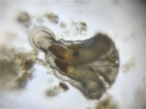
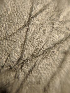
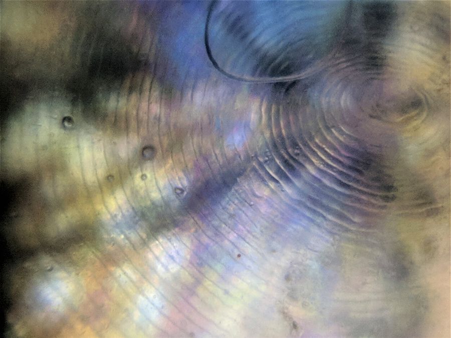
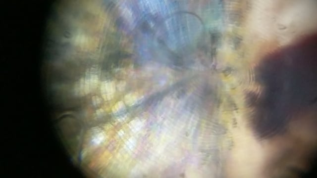
Scales:
One interesting thing I learned is that fish scales have concentric ring patterns, like the rings seen in cut tree trunks! Apparently, the rings (or annuli) tell a story. Counting the number of rings on a scale provides the fish age, and the measuring the spacing between rings is proportional to the growth of the fish.
Now that I know this, next time I encounter some fish scales I want to try a method like this to really try and approximate its age.
Also, you can see some rainbow colors/iridescence, which I believe is from light being diffracted by the rings?
View in Media Gallery
Annuli of piranha scale
Blood:
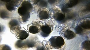
As shown in the gallery of the slides earlier, I took a few drops of the piranha’s blood with a pipette and isolated them into the wells of a paper slide. I was excited to see fish red blood cells because they have some significant differences compared to human red blood cells. Specifically, fish erythrocytes are oval in shape and are nucleated! Being aware of this going in, I hoped to capture an image of the nuclei through a foldscope.
In the video below you can see a thick portion of the blood flowing. (You can also hear me asking for Jim’s phone to image with haha. His phone has a better camera with much higher zoom, which would have helped to really get closer to individual cells.) Alas, I did not catch him or his phone, but on the cropped image below is the best my phone could do. If you look closely, you can see nuclei in the center of the oval-shaped cells.

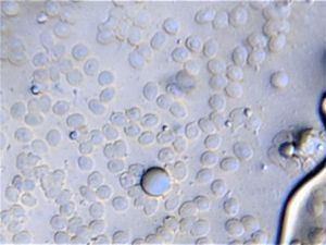
Piranha erythrocytes
Tooth
How could we not? Here is a quick video of the piranha’s tooth, extracted with the foldscope kit’s scissors. As the tooth is pretty opaque, you cannot see much, but around the edges there is some nice definition. For a fun fact, the word “piranha” literally translates to “tooth fish” in the Brazilian language Tupí.
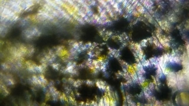
Pectoral Fin
Here is a look at the tissue of the pectoral fin. Just beautiful, brilliant colors and markings.
Here is a look at the tissue of the pectoral fin. Just beautiful, brilliant colors and markings.
Eye
And finally, Jim and I half-joked “What if we look at the eye?” … When I actually attempted to cut it out, not as funny. It was quickly obvious I had no idea what I was doing, as I poked and prodded the goopy socket. It reminded me of cow eye dissections from biology classes of past. A man from the community noticed and offered to help. Immediately he made a cut.
And finally, Jim and I half-joked “What if we look at the eye?” … When I actually attempted to cut it out, not as funny. It was quickly obvious I had no idea what I was doing, as I poked and prodded the goopy socket. It reminded me of cow eye dissections from biology classes of past. A man from the community noticed and offered to help. Immediately he made a cut.
Precise. Perfect. Isolating the eye effortlessly. It dawned on me that the skill made sense, this is a fishing community, of course. But it was simply impressive to see.
Ironically, once the eyeball was extracted, it sat untouched in a fleshy heap. The man who cut it out and I looked at each other and laughed. We didn’t want to foldscope the thing any longer. It was a mess! We just smiled and agreed yeah maybe we got a little carried away in the excitement.
… But later, honestly, I thought about the fish’s iris and had some regrets. I should have at least given it one look.
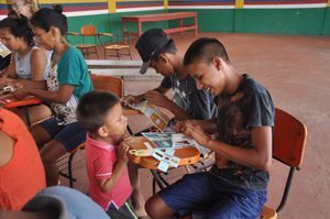
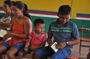
Also, a last note for that dissection video, notice how Judy (Jim’s mom) just casually strolls by?! A woman, who hadn’t left her hometown for the majority of her life, is glancing at a piranha dissection as if it was commonplace. Haha, I loved that. The little boy was also an active spectator throughout the workshop, eventually guided by us all on how to build and use a foldscope of his own.
Ironically, once the eyeball was extracted, it sat untouched in a fleshy heap. The man who cut it out and I looked at each other and laughed. We didn’t want to foldscope the thing any longer. It was a mess! We just smiled and agreed yeah maybe we got a little carried away in the excitement.
… But later, honestly, I thought about the fish’s iris and had some regrets. I should have at least given it one look.
Also, a last note for that dissection video, notice how Judy (Jim’s mom) just casually strolls by?! A woman, who hadn’t left her hometown for the majority of her life, is glancing at a piranha dissection as if it was commonplace. Haha, I loved that. The little boy was also an active spectator throughout the workshop, eventually guided by us all on how to build and use a foldscope of his own.
—
This workshop was an awesome experience, from both the scientific and human perspective. As I think back to it now, I’m not sure I’ll find the words to adequately explain how it struck me– but it did, and I’d like to at least include a nod toward that.
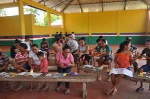
I was thrilled to be able to sit at their table.
I hope I get the chance to return, and eventually, in turn, share more of what I learn with my community.
Until next time!
Rebecca
This workshop was an awesome experience, from both the scientific and human perspective. As I think back to it now, I’m not sure I’ll find the words to adequately explain how it struck me– but it did, and I’d like to at least include a nod toward that.
I was thrilled to be able to sit at their table.
I hope I get the chance to return, and eventually, in turn, share more of what I learn with my community.
Until next time!
Rebecca
Sign in to commentNobody has commented yet... Share your thoughts with the author and start the discussion!
 0 Applause
 0 Comments