Posterior Spircale of Chrysomia species
 Nov 27, 2018 • 3:13 AM UTC
 Unknown Location
 140x Magnification
 Microorganisms
Meignanalakshmi Sundaram
I am Dr.S.Meignanalakshmi, working as Professor, at the Directorate of Centre for Animal Health Studies, TANUVAS, Chennai-51. Working on Foldscope project on "Foldscope for diagnosis of Rumen Acidosis and parasitic infections in cattle" sanctioned by DBT
66posts
8comments
1locations
View in Media Gallery
DBT Foldscope scheme “Foldscope for diagnosis of rumen acidosis and parasitic infections in cattle”
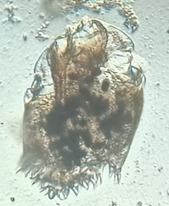
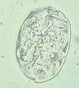
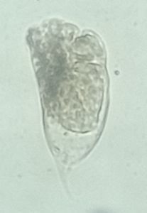
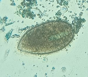
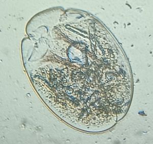
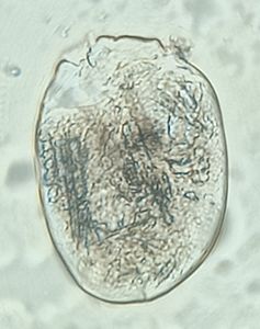
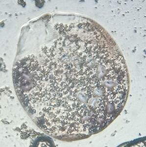
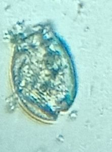
Here’s a beautiful and detailed picture of the processed Posterior Spiracle of a Chrysomya larvae which was collected from an infected wound of a cow.
The steps involved are:
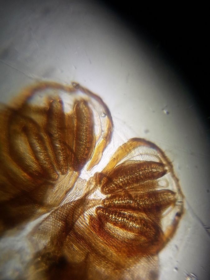
The larvae was boiled in 10% NaOH for about 5 mins and was thoroughly rinsed in water. It was then dehydrated for 2 – 5 mins in increasing concentrations of alcohol ( 70%, 90% and 100%). The larvae was then kept in a clearing agent ( carbolic acid). The cleared larvae was then mounted in DPX mountant and examined under foldscope. The posterior spiracle of Chrysomya larvae (Figure 1) has a pair of more or less circular spiracles with 3 longitudinal slits (b) , incomplete circumference (a) and no button (c).
Here’s a beautiful and detailed picture of the processed Posterior Spiracle of a Chrysomya larvae which was collected from an infected wound of a cow.
The steps involved are:
The larvae was boiled in 10% NaOH for about 5 mins and was thoroughly rinsed in water. It was then dehydrated for 2 – 5 mins in increasing concentrations of alcohol ( 70%, 90% and 100%). The larvae was then kept in a clearing agent ( carbolic acid). The cleared larvae was then mounted in DPX mountant and examined under foldscope. The posterior spiracle of Chrysomya larvae (Figure 1) has a pair of more or less circular spiracles with 3 longitudinal slits (b) , incomplete circumference (a) and no button (c).

View in Media Gallery
Figure 1: Posterior spiracle of Chrysomya species
Sign in to commentNobody has commented yet... Share your thoughts with the author and start the discussion!

 0 Applause
 0 Comments