Helminth Eggs
 Mar 24, 2019 • 8:26 AM UTC
 Unknown Location
 140x Magnification
 Microorganisms
Meignanalakshmi Sundaram
I am Dr.S.Meignanalakshmi, working as Professor, at the Directorate of Centre for Animal Health Studies, TANUVAS, Chennai-51. Working on Foldscope project on "Foldscope for diagnosis of Rumen Acidosis and parasitic infections in cattle" sanctioned by DBT
66posts
8comments
1locations
View in Media Gallery
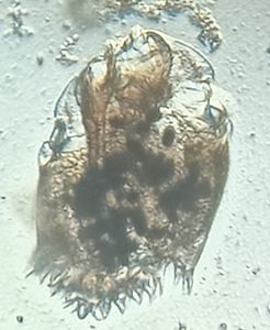
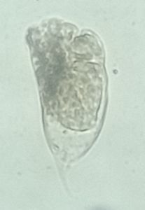
Helminths are large multi-cellular permanent parasitic worms, which can be seen with our naked eyes. Knowledge of various species of helminth affecting the cattle will give an idea about the nature and intensity of helminthosis in bovine. They live and feed on living hosts. Helminths include Trematodes, cestodes, and nematodes.Trematodes are commonly called as flat worms, cestodes are commonly called as tapeworms and nematodes are commonly called as round worms.
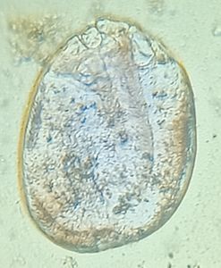
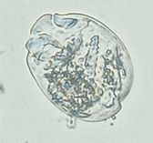
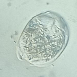
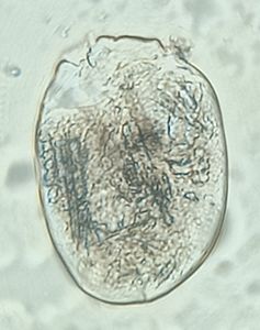
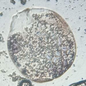
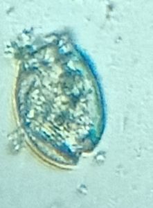
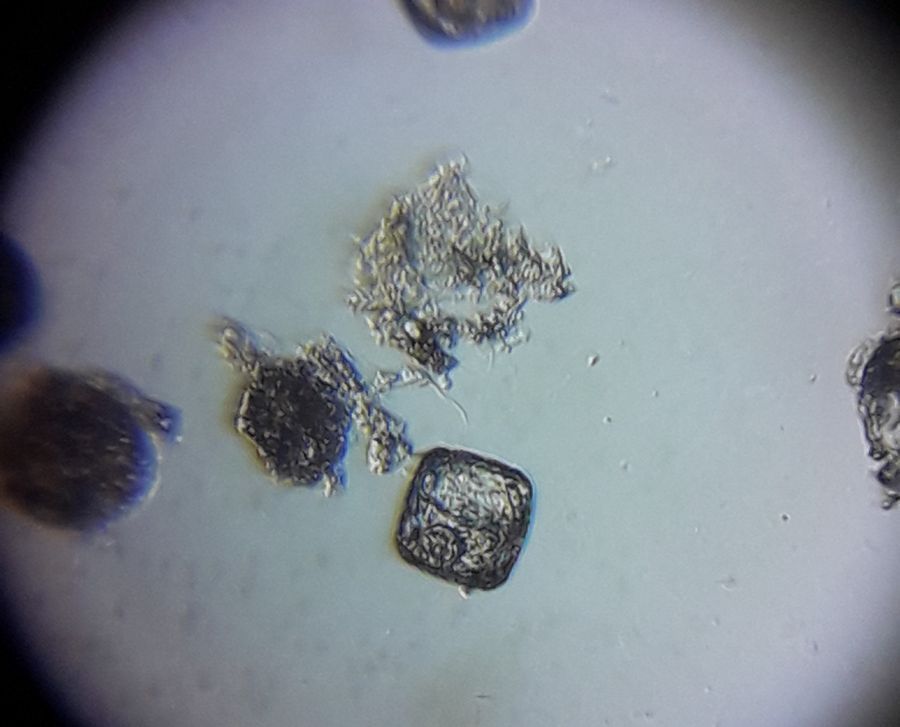
The most common tapeworm in cattle is Moniezia benedeni.
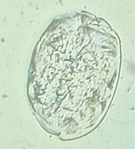
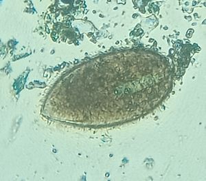
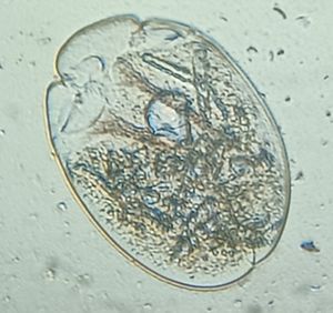
The eggs of Moniezia benedeni are square shaped (Figure 1) and have pyriform apparatus with hexacanth embryo.
Dung samples were collected from cows with the following symptoms: enteritis, rough coat, dehydration, reduced milk yield etc. The different methods of microscopic examination of a faceal sample are: 1. Direct examination 2. sedimentation technique 3. Floatation technique. All the three methods were carried out and eggs could be detected through all the three methods of dung examination by foldscope.
In direct examination method, a small quantity of faeces was placed on a glass slide along with 2-3 drops of water. The faecal sample was thoroughly emulsified with a needle and examined under foldscope.
In sedimentation technique, the following protocol was followed:
A small quantity of faeces was taken. It was emulsified thoroughly with 10-15ml water ( or 0.5% gylcerine in water) and strained to remove all the debris. The filtrate containing the eggs was then centrifuged at 2000 rpm for 1-2 mins. The supernatant was discarded and a drop of the sediment was taken, placed on the slide, covered with a coverslip and viewed under foldscope. In floatation technique, the following protocol is followed:
A small quantity of faeces was taken and emulsified in saturated sodium chloride solution. The thick emulsion of faeces was filled upto one third of the small floatation tube. The tube was filled to its capacity with the same floatation solution. One or two drops more of the solution was added till a convex surface is formed. It was allowed to stand for 15-30 mins by which time all the eggs had floated up. A clean coverslip was placed to the surface of the fluid over the slide and viewed under foldscope. Comparing the three methods, direct examination is the simplest method and it can be used in the field study. But, it is effective only when the helminth infection is high. Sedimentation technique is the most reliable one by which one can recover the eggs of every type of worm- Trematode, Cestode and Nematode. Floatation technique can also be used to detect light infection.
The most common tapeworm in cattle is Moniezia benedeni.
The eggs of Moniezia benedeni are square shaped (Figure 1) and have pyriform apparatus with hexacanth embryo.
Dung samples were collected from cows with the following symptoms: enteritis, rough coat, dehydration, reduced milk yield etc. The different methods of microscopic examination of a faceal sample are: 1. Direct examination 2. sedimentation technique 3. Floatation technique. All the three methods were carried out and eggs could be detected through all the three methods of dung examination by foldscope.
In direct examination method, a small quantity of faeces was placed on a glass slide along with 2-3 drops of water. The faecal sample was thoroughly emulsified with a needle and examined under foldscope.
In sedimentation technique, the following protocol was followed:
A small quantity of faeces was taken. It was emulsified thoroughly with 10-15ml water ( or 0.5% gylcerine in water) and strained to remove all the debris. The filtrate containing the eggs was then centrifuged at 2000 rpm for 1-2 mins. The supernatant was discarded and a drop of the sediment was taken, placed on the slide, covered with a coverslip and viewed under foldscope. In floatation technique, the following protocol is followed:
A small quantity of faeces was taken and emulsified in saturated sodium chloride solution. The thick emulsion of faeces was filled upto one third of the small floatation tube. The tube was filled to its capacity with the same floatation solution. One or two drops more of the solution was added till a convex surface is formed. It was allowed to stand for 15-30 mins by which time all the eggs had floated up. A clean coverslip was placed to the surface of the fluid over the slide and viewed under foldscope. Comparing the three methods, direct examination is the simplest method and it can be used in the field study. But, it is effective only when the helminth infection is high. Sedimentation technique is the most reliable one by which one can recover the eggs of every type of worm- Trematode, Cestode and Nematode. Floatation technique can also be used to detect light infection.
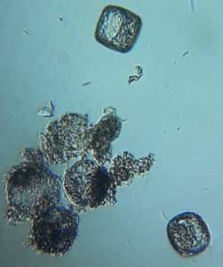
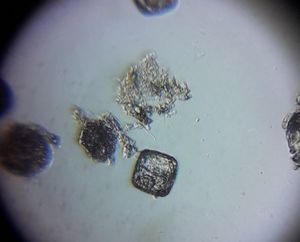
Figure 1: Eggs of Moniezia benedeni.
Sign in to commentNobody has commented yet... Share your thoughts with the author and start the discussion!

 0 Applause
 0 Comments