eggs of Strongyloides species
 Sep 24, 2018 • 2:33 AM UTC
 Unknown Location
 140x Magnification
 Unknown
Meignanalakshmi Sundaram
I am Dr.S.Meignanalakshmi, working as Professor, at the Directorate of Centre for Animal Health Studies, TANUVAS, Chennai-51. Working on Foldscope project on "Foldscope for diagnosis of Rumen Acidosis and parasitic infections in cattle" sanctioned by DBT
66posts
8comments
1locations
View in Media Gallery
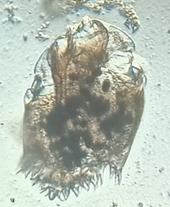
Endoparasites live inside the host. The may either be microparasites such as: blood protozoans (Theileria, Babesia, Trypanosomes) or macroparasites like: Helminths (Trematodes, Cestodes, Nematodes).
Several types of nematodes or roundworms can infect cattle. Although there are many species of worm parasites harbored in the gastrointestinal tract of cattle, only a few target species are clinically and economically important. Strongyloides papillosus is found in the small intestine. Cattle become infected by transmammary, transcutaneous, and oral routes of larval acquisition. Adult roundworms do not multiply in the cattle host. The eggs must pass into the environment to continue the parasite life cycle. The adult life span is only a few months. In order to diagnose a roundworm infection fecal samples were analyzed.
Dung samples were collected from cows with the following symptoms: Green diarrhoea. rough coat, dehydration, reduced milk productivity etc. Sedimentation technique was carried out in order to collect the eggs. The protocol is as follows:
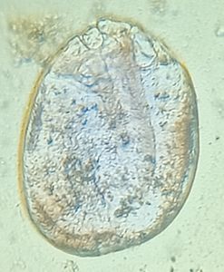
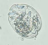
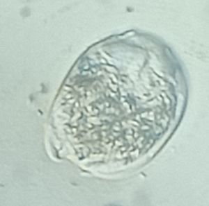
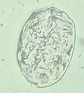
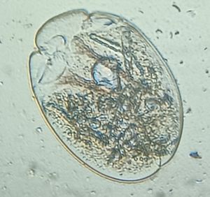
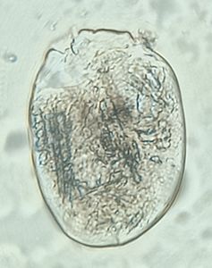
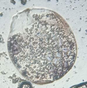
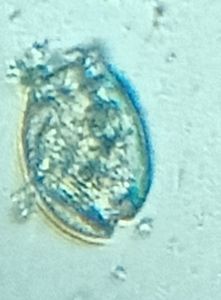
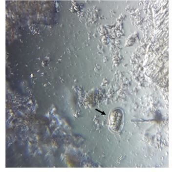
Small quantity of faeces was taken. It was emulsified thoroughly with 10-15ml water ( or 0.5% gylcerine in water) and strained The filtrate containing the eggs were then centrifuged at 1000 rpm for 1-2 mins. The supernatant was discarded and a drop of the sediment was taken and viewed under foldscope. The eggs of Strongyloides were observed to be: (figure 1)
Comparatively small in size oval shape thin shell with both poles blunt well developed embryo inside
Several types of nematodes or roundworms can infect cattle. Although there are many species of worm parasites harbored in the gastrointestinal tract of cattle, only a few target species are clinically and economically important. Strongyloides papillosus is found in the small intestine. Cattle become infected by transmammary, transcutaneous, and oral routes of larval acquisition. Adult roundworms do not multiply in the cattle host. The eggs must pass into the environment to continue the parasite life cycle. The adult life span is only a few months. In order to diagnose a roundworm infection fecal samples were analyzed.
Dung samples were collected from cows with the following symptoms: Green diarrhoea. rough coat, dehydration, reduced milk productivity etc. Sedimentation technique was carried out in order to collect the eggs. The protocol is as follows:
Small quantity of faeces was taken. It was emulsified thoroughly with 10-15ml water ( or 0.5% gylcerine in water) and strained The filtrate containing the eggs were then centrifuged at 1000 rpm for 1-2 mins. The supernatant was discarded and a drop of the sediment was taken and viewed under foldscope. The eggs of Strongyloides were observed to be: (figure 1)
Comparatively small in size oval shape thin shell with both poles blunt well developed embryo inside
View in Media Gallery
Figure 1: Egg of Strongyloides sp
Sign in to commentNobody has commented yet... Share your thoughts with the author and start the discussion!

 0 Applause
 0 Comments