Leaves vs Flowers – How similar and different are they? (BioE 80 Spring 15)
 May 27, 2015 • 12:48 PM UTC
 Unknown Location
 140x Magnification
 Unknown
Iskender Kushan
Learn about the author...
1posts
0comments
1locations
View in Media Gallery
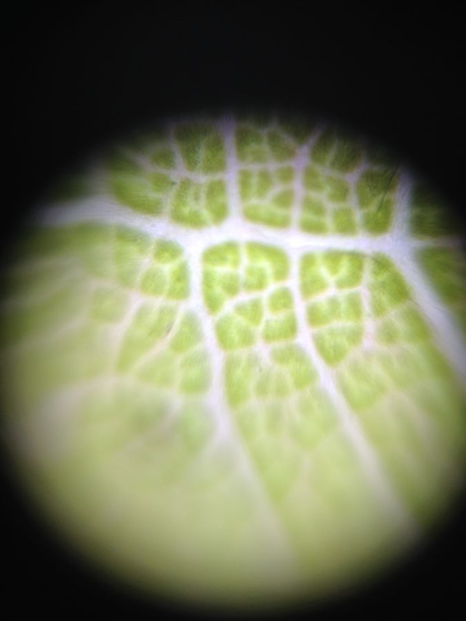
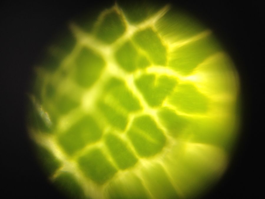
Our exploration of the micro-world began by looking at a leaf. Taken from one of the trees in Stanford’s engineering quad, this leaf was absolutely beautiful under our foldscope. We could see the segments of the leaf, and the vascular bundles (the “veins” of the leaf). Holding our foldscope against the sun allowed the whole leaf to be lit, and produced the best pictures.
View in Media Gallery
Having seen a leaf, we now moved on to other plant parts. How about a flower petal? Would it look similar to a leaf? Would it be as bright as a leaf under the sun?
View in Media Gallery
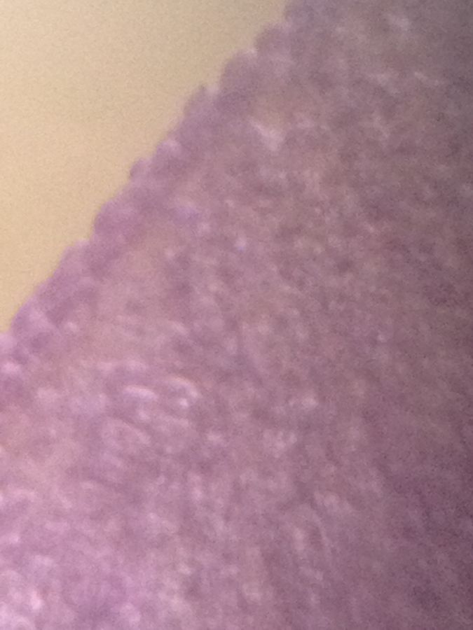
Here is our picture of a purple petal. There were no thick vascular bundles like the leaf on the petals, but this was also something we could see with our own eyes. Yet, the fact that we couldn’t see any was surprising. The petals also did not shine that well under the microscope. A leaf has evolved to perform photosynthesis, so it makes sense that it is thin – that way even the cells embedded most deeply within the leaf would receive sunlight.
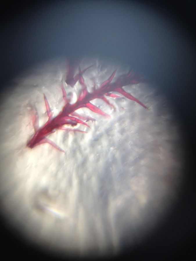
The petal’s internal structure was too small and detailed for our foldscope to resolve, therefore we turned our attention to other parts of the flower. We looked at the stamen, the long and thin part of the flower that is responsible for spreading pollen.
The petal’s internal structure was too small and detailed for our foldscope to resolve, therefore we turned our attention to other parts of the flower. We looked at the stamen, the long and thin part of the flower that is responsible for spreading pollen.
View in Media Gallery
We were able to see the structure of the stamen to our delight. You can also see the barely-resolved pollen as tiny spheres. They are located at the spikes of the stamen, which attach themselves to insects and make it easier for the pollen to stick to the insects. Genius! The fact that the pollen spikes are so reminiscent of Velcro did not escape our attention, and made us appreciate the clever engineers who took inspiration from the nature.
It was amazing how much we could see with our foldscopes -a truly simple, yet brilliant device. Our exploration of the micro-world will not end here 🙂
It was amazing how much we could see with our foldscopes -a truly simple, yet brilliant device. Our exploration of the micro-world will not end here 🙂
Sign in to commentNobody has commented yet... Share your thoughts with the author and start the discussion!
More Posts from Iskender Kushan
No more posts from this author.