Do living flower petals have the same structure as wilting flower petals? (BioE80 Spr2015)
 May 27, 2015 • 1:24 PM UTC
 Unknown Location
 140x Magnification
 Unknown
Ali Hoffer
Learn about the author...
1posts
0comments
1locations
View in Media Gallery
For my exploration I picked a flower petal from a living flower, and then took a flower petal (from the same flower) that was wilting on the soil underneath the plant, and studied both with the foldscope. I expected to see some differences in petal properties and cell organization considering one was dying and the other was still alive. Analyzing both, however, I could not find any differences in cell organization. Both petals appeared to be arranged of small, circular cells packed closely together. What I did notice, however, was that more light appeared to shine through the wilting flower sample, which I presume to be true because it is thinner, and less healthy than the living flower petal. Concluding this exploration there are still a few unknowns and unanswered questions. Namely I do not know how long ago the pedal fell off the flower, so I do not know if the pedal was truly that dead. This could be why the wilting petal and healthy petal looked so similar in structure. Had I conducted this experiment in a more controlled environment wherein I used the foldscope to analyze a living petal, and multiple petals that had fallen off the flower/began to wilt at recorded times, I may have seen different results.
View in Media Gallery
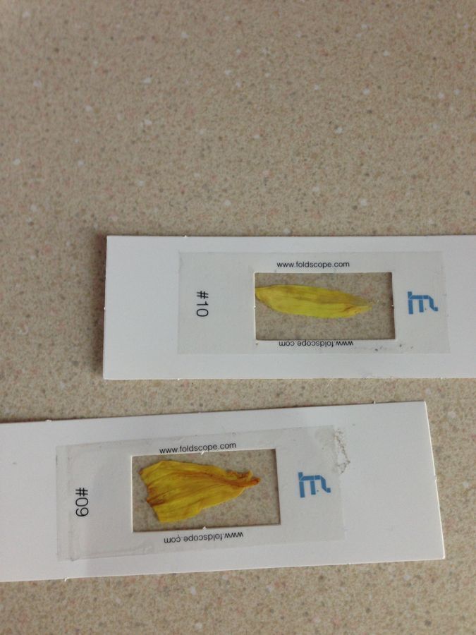
Two Foldscope Slides
Top: Healthy Petal
Bottom: Wilting Pelting
Top: Healthy Petal
Bottom: Wilting Pelting
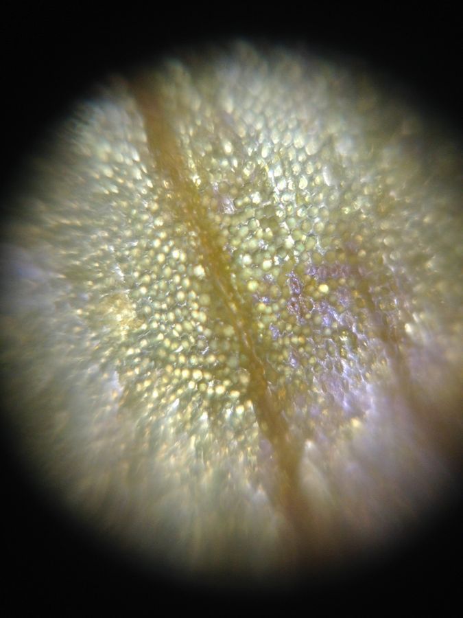
View in Media Gallery
Image of Fresh Petal
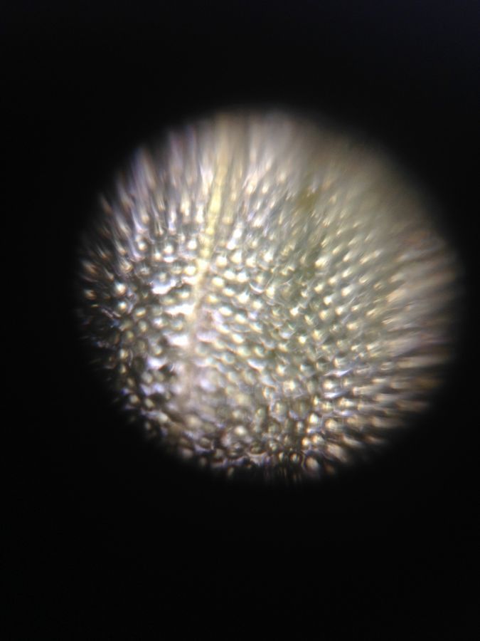
View in Media Gallery
Image of Wilting Petal Explorer: Ali Hoffer
Sign in to commentNobody has commented yet... Share your thoughts with the author and start the discussion!
More Posts from Ali Hoffer
No more posts from this author.